
SUMMER 2008 CONTENTS
Home
Good as Gold?
New drug approvals ebb; doubts over testing's gold standard grow
Q&A with Katie Couric
Standing up for cancer research
Breath of Hope
Lifeline or gamble? Sometimes a clinical trial is both
Just Another Lab Rat
The human subjects trade is booming, largely without oversight
Fixing Trial Tribulations
Solutions from Stanford
A Spoonful of Sugar Pills
Why nothing really is something, and in some ways is better than anything
Banding Together
Minds of all kinds join to hasten discoveries of new medical treatments


Chromosome break
One of the more intriguing workhorses of the cell, a protein conglomerate called telomerase, has in its short history been implicated in some critical areas of medicine including cancer, aging and the health of stem cells.
Gary Taxali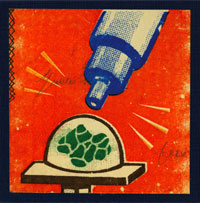
But until now, scientists have been unable to find more than two of the proteins that make up this complex.
Researchers at the School of Medicine have identified two additional proteins that make up the massive telomerase complex and have a lead on several more. This is the first significant step toward understanding the makeup of telomerase since 1999. The discovery also provides new targets for cancer treatments.
“It’s so surprising that we are discovering new components of this enzyme almost 10 years after it was discovered,” says senior author Steven Artandi, MD, PhD, assistant professor of hematology. The work was published in the March 21 issue of Cell.
Telomerase is best known for maintaining the cell’s genetic material, the chromosomes. Every time a human cell divides, a copy of the 46 chromosomes goes to each of the two resulting cells. Each replication snips a bit off the protective tips of the chromosomes, called telomeres. Those ever-shortening chromosomes are one reason cells age. The telomeres eventually dwindle down to a length that triggers the cell to stop replicating altogether or die.
Cancerous cells can overcome that life span limitation by making telomerase, which repairs those snipped chromosome ends. Without shortening, the cells can divide forever. Telomerase is normally active in fetal cells, then shortly after birth it is turned off in all cells except normal-tissue stem cells and some immune cells.
Since telomerase’s discovery in cancerous cells in 1994, the idea has been that if a drug could block telomerase, chromosomes in those cancerous cells would eventually grow shorter and the cells would age and die just like any other cell in the body. But without knowing what proteins make up telomerase, it’s hard to design a drug to block it.
In addition to identifying two telomerase components, Artandi’s work also shows that disabling one of them brings telomerase to a grinding halt. Although the work was done in cells in a lab dish, the findings suggest that a drug blocking that protein might be a useful tool against cancer.
Artandi says the next step is to find small molecules that can block that newly discovered protein in cancerous cells. — AMY ADAMS
This study was funded by the National Cancer Institute, the National Institutes of Health, the American Federation of Aging Research/Pfizer and the Medical Scientists Training Program.
New breed set loose
Mandar Muzumdar came west for medical school at Stanford five years ago. The Harvard graduate was among the first to face a revamped curriculum program that was, if anything, more demanding than before.
As he prepared to graduate in June with the class of 2008, he reflected on his medical school experience, noting that it had exceeded his expectations.
“I heard a lot about the flexibility of the program here,” he says, referring partly to the encouragement students receive to pursue research projects as well as clinical training and basic science preparation.
More than Flexible
He says the curriculum “made sure you had a good grounding in basic medical science. But it also gave the opportunity to tread your own path.” In his case, Muzumdar took an extra year to conduct a research project, which meant he graduated in five years rather than four. But it was time well-spent, he adds. “The research environment at Stanford is hard to beat.”
First offered in the fall of 2003, the new curriculum introduced a scholarly concentration in which each student selects a specific aspect of medicine for multiyear study.
Early results suggest the curriculum change has bolstered the medical school’s education program. One indicator is that more students are matching to one of their top choices for medical residency.
Additionally, the core curriculum was updated to make a more logical progression and to better integrate classroom learning with clinical training.
School leaders have continued to fine-tune the curriculum since its launch. There was discussion as to whether students would need five years to get the full benefit of the curriculum’s research and clinical training, but that decision is being left up to individual students.
“We have a packed, rigorous curriculum,” says Neil Gesundheit, MD, MPH, associate dean for advising, who worked on the curriculum reform. “The idea was to allow a four-year as well as five-year plan without a faculty bias as to which was preferable, and leave that to student choice.”
Of the 87 members of that first class under the new curriculum, 21 received their medical degrees in 2007, while 40 graduated this June.
Early results suggest the curriculum change has bolstered the medical school’s education program. One indicator is that more students are matching to one of their top choices for medical residency. In 2008, the match rate was 93 percent for students’ top three choices, and 75 percent for their first choice.
A second indicator is that the scores for the medical board exams that students typically take after two or three years have edged up since the new program began. At other medical schools, scores have often dropped fleetingly after a curriculum change until faculty and students adjust to the disruption, Gesundheit says. At Stanford, there was a seven-point rise, from a range of 225-229 before the overhaul to 232-235 after. (A score of 230 is in the 80th percentile nationally.)
Charles Prober, MD, senior associate dean for medical student education, says the curriculum change has helped further the school’s goal to train physicians “who are leaders in all aspects of medicine, including research, advocacy and patient care. We want all of our graduates to be thoughtful, caring, compassionate and humanistic physicians.” — DONNA ALVARADO
Map quest
Stanford researchers have created the highest resolution map of human genetic diversity to date, providing insight into how groups of people throughout the world are related and adding weight to previous theories that traced human origins to Africa.
Gary Taxali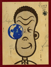
The researchers surveyed 650,000 genetic markers in people from 51 populations to derive the map, providing data that will become a valuable tool in the search for disease-related genes. The work was published in the Feb. 22 issue of Science.
The data confirm earlier work that the vast majority of genetic variation occurs within populations rather than between populations, suggesting that, genetically speaking, race is only skin deep. “Most of the DNA variation we see has nothing to do with what the people who use the term ‘race’ usually mean,” says Marcus Feldman, PhD, professor of biological sciences.
Differences Matter
Unlike previous genetic studies, which surveyed fewer DNA locations and could discern only large population groups, this high-resolution map gives a more detailed view of population diversity. One example is in the Middle East, where the data identify subgroups that had been lumped in a single population. Likewise, the people of China fall into northern and southern groups, whose languages are dramatically different.
The data also give researchers the ability to track human migration, and they support the theory that humans originated in sub-Saharan Africa and left to colonize the rest of the world in several waves.
Perhaps even more important than the study’s findings is what other scientists may be able to learn from the data in the future. The researchers made their data publicly available as soon as they completed the analysis. Although that gave competing groups a head start on projects of their own, senior author Richard Myers, PhD, professor of genetics, says it was the right thing to do. The only stipulation was that other groups allow the Stanford team to publish the first paper using the data, following the precedent set by the public effort to sequence the human genome.
“Like the human genome project, this will enable people to learn important new things about human history and disease,” Myers says.
One place where the data will be useful is in the hunt for disease-related genes. When looking for disease genes, researchers compare groups of people with and without a given disease and look for genetic differences. The data from the Stanford study should help researchers identify which variations are due to genetic heritage, helping them focus on variations associated with the disease. — AMY ADAMS
This study was funded by the National Institutes of Health.
Hypnotic state, seizures abate
It was no way for an 11-year-old to live. For a month the boy had endured daily episodes of uncontrollable jerking and foaming at the mouth, and his physicians at Lucile Packard Children’s Hospital were concerned that the boy had epilepsy. Before starting the boy on a lifetime of antiseizure medications, though, they turned to an unconventional diagnostic tool: hypnosis.
Doctors can pinpoint a seizure’s cause by monitoring brain activity while it’s happening. But while connecting electrodes to a child’s scalp is relatively easy and painless, conducting a “seizure watch” of indefinite length is another matter. Hypnosis shortens the wait.
“Children are highly suggestible and they have great imaginations,” says child psychiatrist Richard Shaw, MD. “We’ve found that if we suggest that they are going to have one of their events while they are in a hypnotic trance, they will usually have one.”
Shaw and Donald Olson, MD, chief of pediatric neurology, tested the procedure on nine children between the ages of 8 and 16. Their results were published online in January in Epilepsy & Behavior. The physicians needed to know whether their seizures were true epileptic events, best treated by medication, or nonepileptic events caused by psychological stress or other neurological problems.
To hypnotize the subjects, Shaw used a combination of deep breathing and progressive muscle relaxation to induce a state of relaxation and focused attention. Then Shaw directed the child to recall the feelings or events that usually precede a seizure. Electrodes on the child’s scalp recorded brain activity during the session.
In eight out of nine cases, Shaw successfully triggered a seizurelike event. After an appropriate monitoring interval, Shaw then directed the hypnotized child to “return” to a favorite place, which stopped the episode. Using this technique, the physicians found that all eight of the subjects’ seizures were nonepileptic.
Stanford is part of a multicenter study seeking treatments for nonepileptic events. For now, Shaw often couples psychotherapy with self-hypnosis lessons to teach children how to avoid the seizures.
“They know that they were able to ‘turn off’ an event during the initial hypnosis, and that gives them confidence to try it themselves,” says Shaw. — KRISTA CONGER
Women’s hearts
Months after joining an exercise group, Valerie Garcia, 48, would stay red in the face, gasping for air, feeling like she was going to pass out, while the rest of her classmates jogged by. No matter how hard or how often she exercised, she was incapable of building any endurance.
Garcia has asthma. Heart problems were the last thing on her mind. But at her doctor’s suggestion she went to the new Women’s Heart Health clinic at Stanford Hospital & Clinics.
Gary Taxali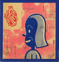
Stress testing showed a possible narrowing in a heart artery, so Garcia had an angiogram. That showed nothing. But after more testing of her heart vessels, she was diagnosed with marked endothelial dysfunction, a form of heart disease that often goes undiagnosed in women.
“I could have easily been dismissed, but they’re specifically interested in women,” says Garcia about her treatment at the clinic. “You can feel it.”
Diagnosing and treating women who might otherwise slide under the radar is the goal of the quickly growing clinic, which opened less than a year ago, says director Jennifer Tremmel, MD, who is also an instructor of cardiovascular medicine. The clinic, launched by the Stanford Cardiovascular Institute, operates one day a week at Stanford Hospital and twice a month at a Monterey clinic, and has about 150 patients.
In step with a national trend to set up women’s cardiology programs, the clinic reaches out to women, whose heart health is overlooked for a variety of reasons — less aggressive care, differing risk factors and gaps in research.
While women are generally more likely to worry about breast cancer, cardiovascular disease kills almost twice as many American women as all cancers put together. It’s the largest single cause of mortality among women, accounting for 38 percent of all deaths among females, according to the American Heart Association.
And heart disease in women is consistently misdiagnosed and goes untreated. Women historically have been left out of scientific studies, though that’s beginning to change.
“We’re finding it just doesn’t work to treat women like men using data derived from men,” Tremmel says. — TRACIE WHITE
Fatal attraction
Four-year-old Braden Eberle was worried. “Mom, I swallowed something,” said the San Jose boy. His mother, Jill, reassured him when she learned that it was just a tiny magnet that had slipped loose from a construction-type toy. But the next day, he swallowed another.
Robert Dicks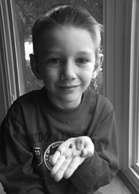
“I didn’t think anything of it at first,” recalls Jill Eberle. But the next day, Friday, Braden began to complain of an intermittent stomachache severe enough to wake him from sleep. On Saturday morning, Eberle took her son to the emergency room — purely as a precautionary measure. “I thought it was probably the flu, but I couldn’t stop thinking about the magnets,” Eberle says.
“Braden didn’t really look that sick,” agrees Lucile Packard Children’s Hospital pediatric surgeon Sanjeev Dutta, MD, who evaluated Braden in April 2007. “But when I heard he’d swallowed two magnets at two different times, I became concerned.” X-rays revealed that the powerful rare-earth magnets had snapped together in Braden’s intestinal tract and were pinching the delicate tissue. Braden needed immediate surgery.
Within two hours, the surgery was over. Dutta used minimally invasive laparoscopic techniques to remove the magnets through three small incisions, and Braden recovered quickly.
Dutta describes the case in a study published in the February issue of the Archives of Pediatric and Adolescent Medicine as a cautionary tale for other physicians. The report urges clinical vigilance and early surgical consultation when magnets are swallowed — even if the child exhibits few symptoms of distress.
“These rare-earth magnets are so much more powerful than the magnets we used to play with as kids. Even one magnet can cause a problem if the child has swallowed something else made of metal.”
Many of the magnets in toys made today contain neodymium, a metal with an unusually strong magnetic force. “These rare-earth magnets are so much more powerful than the magnets we used to play with as kids,” says Dutta. “Even one magnet can cause a problem if the child has swallowed something else made of metal.”
Intestinal tissue pinned between the objects can disintegrate, causing an infection or digestive problems. Also, the affected length of intestine can become twisted, cutting off the blood supply and killing that portion of the bowel.
Several types of toys and games with the magnets have been recalled by the Consumer Product Safety Commission.
“I can’t believe they use these magnets in children’s toys,” said Eberle, who has banned all such magnets from her house. That is, all but two — the two Dutta removed from Braden’s intestine. Those she keeps as a reminder of what could have happened. — KRISTA CONGER