


Turn on relief
Brian Rea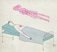
It’s true what the song says: Love is the drug, especially if you’re in pain. The intense feelings of love provide an amazingly effective balm, says professor of anesthesia Sean Mackey, MD, PhD, senior author of an Oct. 13, 2010, report in the journal PLoS ONE about this discovery.
The researchers recruited 15 undergraduates (eight women and seven men) for the study. “We specifically were not looking for longer-lasting, more mature phases of the relationship,” says Mackey. “We wanted subjects who were feeling euphoric, energetic, obsessively thinking about their beloved, craving their presence. We posted fliers … and within hours we had undergrads banging on our door.”
Each was asked to bring in photos of the beloved and photos of an equally attractive acquaintance. The researchers then successively flashed the pictures before the subjects, while heating up a computer-controlled thermal stimulator placed in the palm of their hands to cause mild pain. At the same time, their brains were scanned in a functional magnetic resonance imaging machine.
“One of the key sites for love-induced analgesia is the nucleus accumbens, a reward center for opioids, cocaine and other drugs of abuse. The region tells the brain that you really need to keep doing this.”
The undergraduates were also tested for levels of pain relief while being distracted with word-association tasks such as: “Think of sports that don’t involve balls.” Scientific evidence has shown that distraction causes pain relief, and researchers wanted to make sure that love was not just working as a distraction from pain.
Results showed that love and distraction reduced pain equally, and they did so much more effectively than concentrating on the photo of the attractive acquaintance. Interestingly, the two methods of pain reduction used very different brain pathways.
“With the distraction test, the brain pathways leading to pain relief were mostly cognitive,” says co-author Jarred Younger, PhD, assistant professor of anesthesia. “The reduction of pain was associated with higher, cortical parts of the brain. Love-induced analgesia is much more associated with the reward centers. It appears to involve more primitive aspects of the brain, activating deep structures that may block pain at a spinal level — similar to how opioid analgesics work.
“One of the key sites for love-induced analgesia is the nucleus accumbens, a reward center for opioids, cocaine and other drugs of abuse. The region tells the brain that you really need to keep doing this,” Younger adds.
“This tells us that you don’t have to just rely on drugs for pain relief,” says another co-author, Arthur Aron, a professor of psychology at State University of New York at Stony Brook. “People are feeling intense rewards without the side effects of drugs.” — Tracie White
The study was funded in part by the Chris Redlich Pain Research Fund.
A gene test fails
A genetic marker touted as a predictor of coronary artery disease is no such thing, according to a massive international study led by Stanford researchers.
The study analyzed the data from more than 17,000 patients with cardiovascular disease and 40,000 others to assess whether carrying a particular variant of the KIF6 gene indicated a greater risk for coronary artery disease. The disease can lead to chest pain as well as heart attacks, which are often fatal.
The study, published online Oct. 7, 2010, in the Journal of the American College of Cardiology, found essentially no association between the gene variant and the risk of coronary disease. “This study puts the nail in the coffin,” says Tom Quertermous, MD, professor of cardiovascular medicine and the study’s senior author. “This is such a big study — if there was a significant association between this variant and coronary disease, we would have found it.”
Celera Corp., which pioneered the mapping of the human genome, owns the assay and currently performs the majority of the testing services.
Previous studies of the variant were less conclusive because they were based on fewer patients with coronary artery disease, says the new study’s leader, assistant professor of medicine Themistocles Assimes, MD, PhD. These earlier studies had suggested a 22 to 55 percent greater risk for those who had the variant. “We are showing that the additional risk is almost certainly nil in subjects of European ancestry. If it is not nil, it is at most 2 percent,” Assimes says.
The study pulled together data from research groups around the world that have genetically fingerprinted individuals with coronary disease as well as subjects with no known disease. Most of the data were from people of European descent, but a lack of association was also noted in a smaller number of subjects of non-European ancestry. The Stanford researchers’ co-authors include more than 130 scientists, clinicians and administrators at over 70 research organizations in Europe and North America.
The study offers good news to patients whose KIF6 test result had indicated they were at risk for heart attacks. “They don’t need to worry so much,” Quertermous says. “If they are on medications strictly because of their KIF6 test result, they should ask their doctor to reconsider the need for these medications.”
The finding’s larger message is that more caution is warranted when using genetic markers to guide health care. “We know from previous experience that a positive association between a genetic variant and a common disease, such as coronary disease, needs to be consistently observed in many human population studies before it can be believed,” says Assimes. — Rosanne Spector
The data collections used in this study were supported by more than 30 institutions including government and nonprofit agencies and the following companies: Astra Zeneca, Berlin Chemie, Boots Healthcare, deCODE genetics, Glaxo-Smith-Kline, McNeil Pharma, MSD Sharp & Dohme and Pfizer.
Brain gain
Brian Rea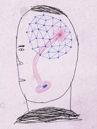
Scientists have found single protein that directs the growth of blood vessels into brains — a discovery that could help enhance blood vessel growth to fight stroke, or choke it off to starve brain tumors.
Calvin Kuo, MD, PhD, associate professor of medicine, and his team discovered the protein’s role in studies of brain development in mouse embryos, which as mammals share many biological features with humans. Kuo is the senior author of the research, published Nov. 12, 2010, in Science.
When the researchers started the experiment, led by Frank Kuhnert, PhD, a research associate in Kuo’s lab, they knew the protein, called GPR124, played a part in blood vessel develop-ment, but they didn’t know what it was.
What they did know was that the protein is a member of a family of proteins called G-protein-coupled receptors that span the membrane that covers cells. Each receptor has a protein partner called a ligand that is secreted into the spaces between cells — usually by a different cell. When a ligand binds to its receptor, it causes a cascade of events within that cell. In this way, the ligand allows cells to “talk” to one another across distances to coordinate many aspects of development and metabolism.
The researchers began by looking to see where in an adult mouse the receptor was normally expressed. They discovered that GPR124 is found almost exclusively on the endothelial cells of the brain and the central nervous system. (Endothelial cells line blood vessels throughout the body and help blood flow more smoothly.) When the researchers bred mice lacking the ability to express GPR124, they died as embryos after about 15 days of gestation. Looking at cross-sections of their brains, it was easy to see why.
“These embryos did not have any blood vessels entering their forebrains or developing spinal cords at all, and the effects were very specific for the nervous system since all other organs had normal blood vessel development,” says Kuo.
In contrast, control mice embryos, with normal expression of GPR124, had already begun developing brain blood vessels after about 11 days.
In the future, the researchers plan to use mice in which they can toggle the expression of GPR124 on and off to examine its role in brain tumor development and stroke. They also hope to learn more about whether GPR124 is involved in the formation of the blood-brain barrier.
“There are a tremendous number of disorders that could be affected by GPR124 expression,” says Kuo. “We’re excited to begin those studies.” —Krista Conger
The research was supported in part by the Stanford Center for Children’s Brain Tumors, the National Institutes of Health, American Heart Association, U.K. Medical Research Council, Brain Tumor Society and Goldhirsh Foundation.
Picking winners
Brian Rea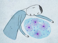
Researchers have found a way to predict within just two days after a human embryo’s fertilization whether it will develop successfully. Since two-thirds of all fertilized eggs fail to make it, the technique could be put to good use, especially at in vitro fertilization clinics.
“It completely surprised me that we could predict embryonic fate so well and so early,” says Renee Reijo Pera, PhD, the senior author of a paper about the technique, published Oct. 3, 2010, in Nature Biotechnology. Time magazine named the discovery one of the 10 medical breakthroughs of 2010.
Professor of obstetrics and gynecology Reijo Pera and her team conducted their study on 242 frozen, one-cell human embryos from an Illinois in vitro fertilization program. When the clinic closed in 2002, the patients gave their consent for their embryos to be used in research.
One of the research team’s aims was to reduce the need for multiple transfers. Because fertilization attempts fail so often, most patients try to increase their chance of success by having more than one embryo transferred into their womb at a time. Yet, multiple transfers lead to other problems. If more than one embryo develops successfully, chances of miscarriage are higher. If a woman has a selective abortion to reduce the number, she improves the survival odds for those remaining — but most women would prefer to avoid such a choice.
Another goal was to cut down on the time embryos spend growing in culture. Nowadays, clinicians usually grow the embryos in culture for three to five days and then pick those that look healthiest to implant or freeze for later use. But this method doesn’t work very well, and concerns are mounting that during culture genetic changes accumulate that can harm the fetus.
The researchers used time-lapse video and computer software they created to track the development of a subset of the embryos from the Illinois clinic — which were ideal for this particular study because they had been cultured for less than a day before being frozen. They followed the embryos through the development of a hollow ball called a blastocyst, which typically occurs within five to six days after fertilization. A blastocyst is usually an indication of a healthy embryo.
They found that 38 percent formed normal-looking blastocysts — about the same proportion that would be expected to be successful in normal pregnancies. Because they had tracked the embryos’ development so closely, they were then able to go back and identify specific parameters that were associated with successful blastocyst formation, among them the amount of time leading up to the embryos’ first division from one cell into two, as well as how long that division took.
If an embryo’s development fit certain parameters, it had a 93 percent likelihood of developing successfully into a blastocyst.
As part of the project, the researchers created an automated algorithm for clinical use that could assess these time-lapse microscopy videos and determine with high accuracy which of these very early embryos would be successful. Stanford has licensed the technology exclusively to Auxogyn Inc. Reijo Pera and the other co-authors of the manuscript own or have the right to purchase stock in the company. — Krista Conger
The research was funded by an anonymous donor, the March of Dimes and the Stanford Institute for Stem Cell Biology and Regenerative Medicine.
Not safe yet
Since a 1999 Institute of Medicine report sounded the alarm about high medical error rates, most U.S. hospitals have made changes in operations intended to keep patients safer. But a look at 10 hospitals’ safety records reveals bad news: Over a recent five-year period, no decreases in patient harm were found at these randomly selected hospitals in North Carolina, a state that has shown a particularly strong commitment to patient safety.
“Our findings are a call to action for the health-care system. We need a nationwide strategy for reducing harm from medical care,” says Paul Sharek, MD, an associate professor of pediatrics, and co-author of the report.
The research was published Nov. 25, 2010, in the New England Journal of Medicine. The study’s lead author is Christopher Landrigan, MD, assistant professor of pediatrics and of medicine at Harvard.
“Our findings are a call to action for the health-care system. We need a nationwide strategy for reducing harm from medical care.”
To perform the study, the team used the Institute for Healthcare Improvement’s Global Trigger Tool. Trained investigators scanned patients’ charts for “trigger” events that suggested harm had occurred. For instance, a prescription for the anti-opioid drug naloxone could suggest an overdose of morphine or a related opioid medication. When reviewers find such an event, they examine the patient’s entire medical record to look for evidence of harm.
The reviewers used this method to examine medical charts from 2,341 randomly selected hospital admissions at 10 randomly selected hospitals in North Carolina between January 2002 and December 2007. The analysis turned up evidence of 588 instances of harm to patients. More than 80 percent of the harms identified were temporary. About half of the temporary harms prolonged the patient’s hospital stay.
Most of the harms were minor or reversible but some were more serious: 50 were classified as life-threatening, 17 incidents resulted in permanent harm to a patient and 14 deaths were attributed in whole or in part to medical errors.
Total harm rates remained the same throughout the study, at about 25 harms per 100 hospital admissions. Separate analysis of different types of harms — more versus less severe and preventable versus non-preventable — did not uncover any subtypes of harm that changed during the study. However, the study did not have the statistical power to evaluate changes in the rate of the most serious harms.
The findings beg the question: Are safety measures useless? Sharek says no. “Implementation of best practices shown to improve patient safety is very difficult and takes time,” Sharek says. Adding to the challenge is the paucity of evidence-based best practices identified in the medical literature for hospitals to implement. More research is needed to separate useful safety interventions from those that do not reduce medical errors, Sharek says. — Erin Digitale
The research was funded by a grant from the Rx Foundation and by funds from the Institute for Healthcare Improvement.