
SPRING 2014 CONTENTS
Home
Fresh starts for hearts
Cardiovascular medicine looks to stem cells for answers
Hiding in plain sight
A high-cholesterol gene
A change of heart
A conversation with Dick Cheney
Switching course
Untangling a birth defect decades later
Dear Dr. Shumway
A boy, two frogs and an airmail letter
Easy does it
Aortic valve replacement without open-heart surgery gains ground

DOWNLOAD PRINTABLE
ISSUE (PDF)
Special Report
Switching course
Untangling a birth defect decades later
By Julie Greicius
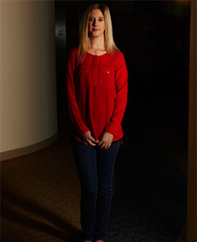
Photograph by Colin Clark
When people ask 24-year-old Brooke Stone about her chest scar,
she gives them the simplest answer she can: “I tell them I was born with my heart attached backwards.”
As a newborn in 1988, Brooke was diagnosed with a congenital heart defect known as dextro-transposition of the great arteries, and underwent a complex surgery to correct her blood flow.
Stone’s doctors knew that this surgery would probably not be her last. Of the 40,000 children in the United States born with congenital heart disease each year, 50 percent will require at least one major surgery, according to the Children’s Heart Foundation. Of those, almost all will require regular follow-up care, with many needing one or more additional surgeries because of later-life complications. Those whose lives are saved by early surgical intervention will never be entirely free of the risks of their condition.
The challenge today is to ensure that post-surgical patients survive long enough to benefit from advances in care that are evolving as patients age. Surviving means receiving ongoing monitoring and care — which only about half of adolescent and adult patients currently receive — allowing doctors to intervene before patients suffer irreversible cardiac damage.
Some lifesaving advances take decades to achieve. The second procedure that saved Stone’s life evolved over more than half a century, the result of work by five surgeons from five different countries — Sweden, Canada, Brazil, Australia and the United States.
“We waited 24 years for this surgery,” says Stone’s dad. “Twenty-four years.”
Because the heart is so complex, its defects are diverse, with more than 40 different types. Transposition of the great arteries alone has several variations, of which dextro- (or “complete”) is just one. In dTGA, the two main arteries that come out of the heart — the pulmonary artery and aorta — are connected to the wrong chambers of the heart. As a result, blood fails to circulate properly: Instead of circulating from the heart to the lungs, where it gathers oxygen, back to the heart and then out to the rest of the body, it flows in two closed loops. In a heart with dTGA, blood from the lungs flows back to the lungs and blood from the body flows back to the body without ever getting the oxygen it needs.
�I was upset. I mean, I was living a normal life. I was in college. I was fine. I could not understand why they wanted to take a perfectly fine person and do this.�
“Transposition of the great arteries was almost 100 percent fatal before the late 1950s,” says Frank Hanley, MD, director of the Children’s Heart Center at Lucile Packard Children’s Hospital and a professor of cardiothoracic surgery at Stanford. Hanley has been repairing the hearts of infants born with TGA for decades. But, over the years, repair for TGA has evolved dramatically. Patients like Stone, whose lives were saved by an earlier surgical method developed in the late ’50s, may find themselves aging out of that childhood surgery and facing heart failure anew.
Stone’s first surgery — a Senning procedure, named after Åke Senning, the Swedish surgeon who developed it in 1957 — was the most trusted repair available for TGA at the time. It would be years before surgeons would become technologically advanced enough to reliably switch the misplaced arteries to their anatomically correct position. Instead, the Senning procedure was an “atrial switch,” which opened a space in the wall between the heart’s two upper chambers (the right and left atria) and redirected the blood flow using a baffle — a panel constructed out of heart tissue — to guide blood correctly through the transposed arteries and allow oxygenation. “Instead of righting the wrong,” says Hanley, “they added these two wrongs to make a right.”
Sitting with her in a bright café eating lunch, it’s hard to imagine the radiant, healthy adult Brooke Stone as a 4-pound, 10-ounce newborn with skin tinted blue from her lack of oxygen. But for her parents, Barb and George Stone of Paso Robles, Calif., those memories are vivid.
Right after Brooke’s first surgery at University of California-San Francisco, doctors told Barb that Brooke would not survive adolescence without additional surgery. So when Brooke was still a baby, Barb began decades of international detective work, calling hospitals and universities, searching for breakthroughs and doctors who could help her daughter. Each time, she was led back to one of the doctors who, as a young surgical fellow, had assisted in Brooke’s first surgery: Frank Hanley.
By then, Hanley was practicing at Packard Children’s heart center and developing his own lifesaving interventions for congenital heart disease. In the Adult Congenital Heart Program at Stanford, Hanley, a slender, silver-haired man who favors a tweed jacket over his scrubs when he’s not in surgery, also treats many adults — some even in their sixth and seventh decade of life. Some patients have had a problem similar to Brooke’s: a failing right ventricle resulting from an early-childhood Senning procedure — or from a similar atrial-switch named after the Canadian surgeon, William Mustard, who refined it in 1964.
The early Senning and Mustard repairs saved the lives of countless children who did well through their 20s, 30s and sometimes 40s before anyone realized there was a problem: The right ventricle, part of the heart muscle that normally pumps blood at low pressure to the lungs, was never designed to pump at the higher pressure needed to push blood through the whole body. After decades of bearing this burden, the right ventricle could eventually fail. Young adults like Brooke suddenly found themselves with an entirely new, life-threatening heart problem.
“Doctors had three choices for solving this problem,” explains Hanley. “The first was to manage these patients with medications — not a complete cure. The second was heart transplant — an imperfect solution because of limited donors and the potential for organ rejection.” The third, rarest, approach was potentially the most beneficial: undo the original repair and move the originally transposed arteries back to their correct positions.
Switching the arteries back to their correct positions was first successfully performed in newborns by Brazilian surgeon Adib Jatene, MD, in the late 1970s. It proved more challenging in adults.
In a normal heart, the left ventricle does the heavy lifting, pumping blood through the whole body. But for patients with TGA who had the Senning or Mustard procedure as infants, the left ventricle becomes like a strong man who’s been sitting on a couch his whole life, gently pumping blood at low pressure to the lungs. The left ventricle needs training to get back in shape to pump at a pressure strong enough to move blood through the whole body as it was designed to do. “The heart muscle is just like the muscles in your arms and legs,” explains Hanley. “If you tell people who’ve never worked out to go to the gym and bench-press 200 pounds, they probably can’t do it. But if you train them for a year, maybe they can. It’s the same with the heart muscle.”
In the late 1980s, Hanley was the first to follow Australian surgeon Roger Mee, MD, in training a patient’s left ventricle by placing a band around the pulmonary artery. By tightening the band at intervals over the course of 12 to 18 months, the left ventricle would have to work harder to push blood through this smaller opening, increasing its ability to pump at higher pressure. The heart would then be ready to have its old Senning or Mustard repair undone and an arterial-switch performed to fully correct the heart.
“Like a lot of new things,” says Hanley, “there was a flurry of interest and a whole bunch of cutting-edge institutions jumped on board to try it. A lot of those patients didn’t do well, and many surgeons were discouraged by the bad results. So they abandoned it.” But not Hanley. He looked closer. “The idea that everyone who needed the procedure could just be slam-dunked into the arterial switch was wrong,” he says. “We focused on setting rigid criteria for accepting people into the program, and setting up a five-point report card after left-ventricle training to ensure that we were selecting appropriate patients who would have good outcomes.”
Today, Hanley is the only U.S. surgeon still doing the procedure. His careful process, which involves lifelong monitoring and management by a cardiologist — and eventually by Hanley himself — as well as patient selection and rigorous evaluation, is key to his approach. Now, when he carries out these surgeries, the outcome is successful more than 90 percent of the time, proving that the procedure is a viable and even advantageous alternative to transplant.
“My whole life I knew that I had to have something,” says Brooke.
“There was always this inevitable something.” Knowing does make it easier. Many patients with congenital heart disease mistakenly believe that if they’re feeling fine, then their heart must be doing well. And if they believe they’re doing well, then they don’t visit the doctor.
“You go on throughout your whole life feeling normal,” says Brooke. “It almost felt like getting diagnosed with something new.” But, as she discovered, the heart of a person with congenital heart disease may be weakening and experiencing potentially lethal, abnormal rhythms, known as arrhythmias, without any outward signs or symptoms. During the summer of 2012, when her arrhythmia was first discovered, she says, “I felt the best I’ve ever felt in my whole life.”
Luckily, Brooke had been carefully monitored — a critical aspect of care for anyone with congenital heart disease. Each year, Barb made an appointment with the family’s local cardiologist, John Owens, MD, for a complete study of Brooke’s heart function. As the years passed, and Brooke felt healthy and more independent, it became ever harder to justify annual checkups.
“I was upset,” says Brooke. “I mean, I was living a normal life. I was in college. I was fine. I could not understand why they wanted to take a perfectly fine person and do this.”
The desire to lead a “normal” life can be a slippery slope. “For Brooke’s sake,” says Barb, “we always treated her as a normal child. She couldn’t go to the mountains and she couldn’t do sports, and she couldn’t do a lot of things. But nobody knew, unless they looked at her scar and asked. I thought it was important for her to be treated normally by all of her peers. We began to think that way, too, because that’s what happens. And then when a doctor says, ‘We’ve got to go in,’ you think: You’ve got to be kidding. Look at her!”
Yet, Brooke’s parents knew monitoring was vital to her survival. Each year, in addition to the necessary echocardiograms, catheterizations and MRIs, her cardiologist sent her home with a 24-hour monitoring device that would record Brooke’s heart activity as she went about her day.
“It was the fanny pack,” says Brooke’s father, referring to the belt-case that held the 24-hour Holter monitor. “The fanny pack gave it away.” The monitor recorded the life-threatening heart rhythms that were going on as Brooke slept.
So it is with many congenital heart disease patients: Catching the failing heart with monitoring is the often-ignored solution to early intervention and survival. “Catching it at the right time saved my life,” says Brooke.
In Brooke’s case — and with similar patients managed by Hanley — a carefully monitored left-ventricular outflow-track obstruction had narrowed the space in her pulmonary artery and increased pressure from the inside, just as Hanley’s band-tightening procedure would have done from the outside. The result was that Brooke’s left ventricle was already strong enough to pump blood to the body, making Brooke — and patients like her — an ideal candidate for the arterial switch procedure, with no left-ventricle training required. She would, however, need an additional procedure the following year to replace her left ventricular outflow track. This procedure, also performed by Hanley, finally put Brooke’s heart in nearest-to-normal condition — the result she and her family had sought for so many years.
To date, Hanley has managed 36 patients with failing atrial-switch — a number rivaled only by retired surgeon Roger Mee. For 21 of those patients, Hanley completed the atrial-switch takedown and arterial-switch operation, with an overall survival rate of 81 percent. But the overall success rate doesn’t reflect his increasingly positive results: Over the past 15 years, as the criteria for selection and the procedure have evolved, the survival rate is much higher, with the last unsuccessful procedure performed in 1999. Twelve new patients are undergoing pulmonary artery banding. Early in the development of this procedure, two patients died of right ventricle failure before training was complete. Hanley notes that today these two patients would not qualify for the surgery because their hearts had decompensated to an extent that the procedure, he now knows, would be unsafe. These early cases helped guide Hanley to the further-refined, safer criteria he uses today.
Hanley estimates that thousands of adults who had Mustard or Senning repairs in childhood may now need interventions of some kind, though many may not know it.
“During that 25-year period from 1959 to the early ’80s, atrial-switch repair was the only solution available to these patients,” Hanley explains. “This means there are thousands of people alive now with atrial switches in whom this right ventricular failure risk is lurking.”
As the most common birth defect, congenital heart disease kills twice as many kids as all childhood cancers combined. Those who survive depend on their parents to stay ahead of the disease. And yet, “many parents of children with congenital heart disease don’t recognize that their kids need lifelong cardiac care,” says Susan Fernandes, program director of the adult congenital heart program at Stanford, and lead author of a 2011 study on the topic. “It is estimated that more than 50 percent of adults with congenital heart disease are not receiving specialized, adult congenital cardiac care or are lost to follow-up, most falling out of appropriate care before mid-adolescence.”
Those who are monitored are often followed by a primary care doctor who may not know about the potential complications of their condition. This unseen risk points to the larger problem of how to provide the best monitoring and care of survivors of congenital heart surgery.
“There’s a big push among those who care for congenital heart patients in this country to organize adult survivors of congenital heart surgery and bring them into clinics where they can be treated effectively,” says Hanley. Lucile Packard Children’s Hospital Stanford is one of those, with its multidisciplinary adult congenital heart program. This program is a major priority for the institution. Stanford recently recruited George Lui, MD, clinical assistant professor of cardiovascular medicine, as medical director of the program.
The push for more organized care can’t happen soon enough. With resources and clinic directories for patients across the country, the Adult Congenital Heart Association is a leader in this effort.
“It’s a real big timing thing,” says Barb. “And there are so many kids who are left until it’s too late.” Staying on top of the disease gave Brooke treatment choices she may not have otherwise had and allowed her to plan ahead.
Hanley told Brooke she would need surgery within three to five months. She could pick the date, which allowed her to graduate college and enjoy her summer.
Her eight-hour surgery took place at Packard Children’s on Sept. 27, 2012. Expecting a three-week stay, she recovered in nine days and was released.
“She came out of the hospital and smiled,” says Barb. “She looked up at the blue sky and she started crying. That was a moment I will never forget, because I could see the feeling in her, like, ‘I am alive.’”
Today, Brooke is living in Aptos, Calif., with her dog, Maddy, and taking some time to relax and appreciate life. She takes every opportunity to get the word out to young adults living with a congenital heart defect about the importance of appropriate care, giving a talk at the Lucile Packard Foundation for Children’s Health last September.
She’s also doing supervised interval workouts six days a week. And she’s taken up the hula hoop. “It feels great to be using my body to its full potential,” says Brooke.
“I’m looking forward to running,” she says, “and just being able to not have any limitations — living to live instead of living to survive.”
For her parents, getting used to the promise of Brooke’s healing heart is an adjustment. “I still take each day at a time,” says Barb. “As of right now, it’s just hard to believe this is real. It’s the most amazing thing. We’re so grateful.” SM