
FALL 2011 CONTENTS
Home
Game Change
On the verge of a revolution
Inside the labs
Highlighting recent Stanford cancer research
Cancer's biographer
A conversation with Siddhartha Mukherjee
The unexpected
Cancer during pregnancy
After cancer's cured
What's left to heal?
Make your own cancer diagnostic test
It's easier than you think

DOWNLOAD PRINTABLE
ISSUE (PDF)
Plus
Drawn together
Artist and surgeon convey the body’s secrets
By Tracie White
Photography by Leslie Williamson
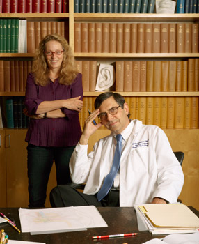
Artist Chris Gralapp and surgeon Robert Jackler have collaborated for the past 20 years on illustrations that depict delicate surgeries of the inner ear and places deep within the skull.
“I counsel you not to cumber yourself with words unless you are speaking to the blind. ... How in words can you describe this heart without filling a whole book? Yet the more you write concerning it, the more you will confuse the mind of the hearer!” — LEONARDO DA VINCI
Every other week on Monday afternoons, a container of colored pencils is placed prominently on the coffee table in the surgeon’s office. Nearby is a sketchpad. Such simple old-fashioned instruments, the pencil and paper. Then the surgeon and the artist enter the room, and the recipe is complete.
Even with the advent of all kinds of advanced technological illustrative and imaging devices, these are still the tools that medical illustrator Chris Gralapp and surgeon Robert Jackler, MD, use to illustrate some of the most technical of microsurgical techniques — hypoglossal-facial anastomosis, middle fossa craniotomy and stapedotomy, among others. Page after page of their drawings already comprise such tomes as Jackler’s Atlas of Skull Base Surgery and Neurotology, which explains the “how-tos” of surgeries deep inside the skull.
And it all begins with a crude pencil sketch. “I’m a terrible drawer,” says Jackler, professor and chair of otolaryngology, who for years has arrived fresh from surgery — removing tumors found in the base of the skull, restoring hearing, repairing eardrums and other problems — and begun describing in vivid detail to Gralapp exactly what he has done inside his patients’ heads. Often he scribbles on paper to help convey his thoughts. “I make my messy sketches,” he says, “and she brings them to life.”
Jackler, together with Gralapp, who has a master’s degree in medical illustration, has published three illustrated books on skull base surgery, and they are now working on a fourth, an exploration of middle ear surgery. The works are also available online, and the pair are going virtual. With help from Nikolas Blevins, MD, associate professor of otolaryngology, and colleagues in computer science, they’re creating enhanced 3-D videos that will allow users to feel the images when they touch them.
The collaboration: Robert Jacklers Sketch (left) of the removal of the tiny stapes bone inside the ear is the basis for Chris Gralapps final illustration.
Their work is inspired by the famous medical illustrators throughout history — Leonardo da Vinci, Andreas Vesalius, Max Brödel — who have used art to describe the structure of the human body and to elucidate medical procedures. “It’s really a natural marriage, this combining of art and science,” says Gralapp, who strives to follow in the footsteps of her heroes — the Renaissance artists who melded scientific understanding of anatomy with artistic skill. “Both work to explain the world.”
Jackler has found that, for him, the use of art is simply the best way to communicate his wealth of experience that few others have. “We’ve spent more than 20 years drawing things that have never been drawn before,” he says.
On a recent Monday, Jackler’s shirtsleeves are pushed up. Gralapp’s long hair is pulled back in a headband, a sketchpad in hand. The surgeon and the artist are reworking the third drawing of what will eventually be about 50 or 60 in a series used to illustrate a single micro-surgical procedure, which takes place in an area the size of the head of a pin. Called a stapedotomy, this surgery is the only way to fix abnormal growth or hardening of the middle ear bone — the stapes — which leads to progressive hearing loss. The procedure involves removal of the arch of the stapes bone, the smallest and lightest bone in the human body, and replacement with a teeny-tiny piston. First performed in 1956 and steadily refined since, it’s surgery on a minute scale.
This particular surgery has been illustrated in other books, but it’s Jackler’s surgical techniques developed over years of fixing problems that will be newly drawn. Just how does the surgeon deal with a slipped crimp? What does the surgeon do if the prosthesis is too long? It’s Jackler’s rare expertise at revision surgery — he’s the guy who gets called in to repair complex surgeries that went wrong — combined with Gralapp’s artistic talent, that create books that fellow surgeons swear by.
“The pictures are so clean they help you walk through the operations mentally before you operate,” says Richard Gurgel, MD, a neurotology fellow at Stanford who specializes in surgeries on tumors of the ear and skull base. He studies Gralapp’s illustrations in the version of Atlas of Skull Base Surgery and Neurotology on Stanford’s website the night before his surgeries, often the removal of benign tumors from cranial nerves.
Other surgeons use the images right inside the operating room. “Before the book went online, surgeons often called from operating rooms and said, ‘I’m looking at your book. Can you clarify a bit?’” Jackler says. Now most study them on the computer, but he still gets the calls.
Gralapp’s goal is to clarify the complex. “As an illustrator I eliminate the unnecessary,” she says.
For the stapes procedure, she’s imagined she’s a nano-sized munchkin who is sitting inside a human ear, and she is drawing the illustrations from that point of view. “It’s a different perspective than what the surgeon sees peering into the ear when he does the surgery,” she says. “But you can’t always see clearly the concept of the surgery from the surgeon’s point of view. This perspective is much easier for people to understand.”
There will always be a role for the artist in science, Gralapp says, as long as there are medical websites out there that need art, as long as there is the complex that needs to be clarified. “We communicate better, far better, through art,” she says.
Final view: These illustrations show a few of the steps involved in placing a tiny piston in the ear after the removal of the stapes.
On this Monday afternoon, Jackler guides her through the stapedotomy.
“This type of microsurgery can be devilishly tricky at times,” Jackler says, looking down at Gralapp’s sketch-in-progress laid out on the coffee table.
“This minuscule bone,” he says, pointing out the stirrup-shaped middle ear bone in Gralapp’s illustration, “the stapes, should be able to move, like a piston.” He uses his hands to show the movement of a piston, a fist pumping against an open palm.
The surgeon’s hands are his greatest tool, and Jackler uses them often when he talks. Gralapp understands what he’s describing almost before he describes it.
Jackler’s obvious excitement about the collaborative process reveals an interest in art beyond the hobbyist. His wife, Laurie, is an artist. Prints of the earless Vincent van Gogh self-portrait have remained prominent at his desk for years. He decorates his walls with 18th- and 19th-century medical illustrations. “We stand on the shoulders of giants,” he says.
Twenty-four years ago, when they first started working together, meeting up at UC-San Francisco — Jackler, a youngish surgeon, and Gralapp, a youngish illustrator — the artist would occasionally join the surgeon in the operating room. But after a few years she stopped coming to watch. They developed their own creative process, and it is one that works best for them here in the quiet of the office with the sketchbook and the pencils.
The two share their own visual language using colors and simple shapes to illustrate surgeries often microscopic in scale. They speak in colors and symbols and the intricacies of difficult-to-get-to anatomical spaces that few in the world have seen up close and personal.
“I think blue would describe this better,” Jackler says. And, “When I think of the tympanic membrane I think of clouds. It’s almost all water.”
Years of practice have honed Gralapp’s interpretation skills, and she quickly brings his words to life. “I’ve been a bit impressionistic here,” she says.
He nods. “That’s beautiful,” he says. Then makes a few more suggestions.
A little more shading, a little bit more shaping, and they’re one step closer to the truth.
E-mail Tracie White

Web Extra
See the surgeon and artist working together