
SUMMER 2010 CONTENTS
Home
Transformers
How teaching hospitals could lead medicine’s metamorphosis
No holes barred
Interest grows in using natural openings for surgery
The healing hand
Putting the physical back in the physical exam
Take the tube
Mass transit for lab samples
Code green
New hospitals blend healing and conservation
On the record
The nation’s health information technology leader on the future of patient data

DOWNLOAD PRINTABLE
ISSUE (PDF)
Special Report
The Stanford Medicine 25
Exam techniques every doctor should know
Photo by Lenny Gonzalez
On a Thursday morning, about 40 internal medicine trainees gather in a Stanford Hospital room to learn what a hand can reveal about a person’s health.
It is the monthly Stanford Medicine 25 session, one in a series of workshops teaching 25 essential techniques for examining patients.
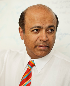
“My worst nightmare is a patient with a diagnosable, treatable disease that I missed because of sloppy technique,” Abraham Verghese, MD, professor of medicine, says in opening the session.
Verghese, the Department of Medicine’s senior associate chair for the theory and practice of medicine, designed the Stanford Medicine 25 with first-year residents (interns) in mind. He hopes that they will perfect a repertoire of hands-on diagnostic skills they can pass along to next year’s crop of interns when they become their immediate supervisors. But the Stanford Medicine 25 sessions have proved popular with second- and third-year residents as well, and the room is full. All trainees have been taught these techniques or know them in theory, but the sessions emphasize hands-on practice.
Each gathering, part of the required curriculum, focuses on a single diagnostic technique and what it can reveal. Verghese uses real patients, standardized patients (actors), and occasionally, as in the session this morning, trainees practice on each other. He also uses simulators, in particular for rectal and scrotal exams, and is enthusiastic about a newly ordered cardiopulmonary simulator — a mannequin displaying normal and abnormal heart impulses and sounds.
Verghese on the techniques
1. Funduscopic exam: Using an ophthalmoscope to examine the fundus (the retina, vessels and nerves in the back of the eye) can help assess the condition of blood vessels throughout the body, diagnose neurologic problems and provide clues to systemic diseases from heart valve infection to AIDS.
2. Pupillary responses: This session covers how the pupils constrict and dilate to light and respond to distant and near vision, as well as the best ways to elicit these findings. The responses can indicate trauma to the eye, and neurological disease and other conditions.
3. Thyroid exam: Palpating the neck to feel the thyroid gland can help diagnose thyroid disease. A nodule can indicate thyroid cancer. Without thorough training, people often feel too high on the neck or place their fingers at an angle that precludes feeling a nodule.
4. Neck veins: Because the jugular veins in the neck go directly to the heart, they can indicate cardiovascular problems. Seeing the neck veins and discerning their pulses takes a practiced eye, good patient positioning, good light and patience. Once it’s seen, the pulse level can be measured and abnormalities identified that can diagnose cardiac conditions such as tricuspid incompetence and complete heart block.
5. Lung: Percussing (tapping) on the chest and sounding out the lung’s boundaries are useful for detecting fluid or pneumonia, particularly in areas without access to radiology equipment and blood testing.
6. Point of maximal impulse and parasternal heave: The PMI is a dime-sized area of the chest, just left of the breast bone, where the beating of the heart can be felt. Heart and lung problems, such as hypertension or cardiomyopathy, create unique PMIs. The parasternal heave is an impulse originating in the heart or large vessels that can be felt with the heel of the hand resting on the left sternum. Though these are crude and simple maneuvers, they reveal much about the heart and can help physicians ask better questions of echocardiograms they order.
7. Liver: This session covers percussion to approximate liver size as well as techniques to feel the liver edge and to feel its surface for nodules and masses. It includes feeling for tenderness in the gallbladder region and signs of gallbladder inflammation.
8. Palpation, percussion of spleen: The spleen is notoriously difficult to feel, yet it is embarrassing to miss an enlarged spleen. When enlarged it is almost always abnormal: It can be a sign of infection, tumor or liver disease. Positioning both the patient and the examiner properly is critical for success.
9. Common gait abnormalities: A person’s walk can indicate nervous system and musculoskeletal problems. The long hospital corridors provide a great opportunity to observe gait abnormalities common in patients with a stroke or with Parkinson’s disease, or peripheral neuropathy (damage to nerves outside the brain and spinal cord) and multiple other conditions.
10. Ankle jerk: This is a natural reflex, a brisk forward movement of the foot, which occurs when a hammer strikes the Achilles tendon above the heel. An absent reflex might suggest nerve damage, but often a reflex is labeled absent only because of incorrect technique (in a bedridden patient in particular). The ankle reflex is almost a metaphor for the Stanford Medicine 25. Being able to elicit this reflex generally means the examiner can elicit the other reflexes, which are easier to bring out.
11. Stigmata of liver disease: The paradox of liver dysfunction is that its signs are found outside the abdomen. These so-called stigmata include spider angiomas (dilated capillaries) on the cheeks, parotid gland enlargement, diminished armpit hair, breast enlargement in a male, islands of redness on the palms and myriad other findings.
12. Internal capsule stroke: An area deep in the brain called the internal capsule is one of the most common sites of stroke. The condition produces a plethora of neurological signs that can be demonstrated, involving cranial nerves, muscles, sensation, reflexes and gait. In this session, the student runs through a series of maneuvers from head to foot that help identify the location of the stroke.
13. Knee exam: The knee is often affected by disease and by trauma. Well-validated means exist for establishing the presence of fluid in the knee and testing for tears in a meniscus or ligament — each test involves a specific physical manipulation, which requires practice.
14. Cardiac second sounds/splitting: The healthy adult has two normal heart sounds (the familiar lub dub), produced when heart valves close. The second sound is actually composed of two separate sounds produced by closure of the aortic valve and the pulmonary valve. Though they close together they become asynchronous after a deep breath. Many variations on this theme — exaggerated splitting or paradoxical splitting or fixed splitting — can speak to specific conditions such as bundle branch blocks or atrial septal defect to name two.
15. Involuntary movements: These range from tremors to much more complex movements. In this exercise, students learn to identify and characterize the types of tremors as well as other involuntary movements termed chorea, athetosis and several more.
16. Hand: Many diseases show signs in the hand, from Down’s syndrome (evidenced by an extra crease in the palm) to certain cancers. The nail is affected by disorders ranging from cystic fibrosis to lung cancer. In this session, students learn to read the hand for everything from nerve disorders to specific finger deformities that in turn predict systemic disease.
17. Tongue: Visually inspecting the tongue for swelling, unusual color or texture can reveal signs of oral cancer, nutritional deficiencies or infection, such as HIV.
18. Shoulder: Like the knee, the shoulder joint is commonly affected by injury and aging. A series of observations and maneuvers can lead the clinician to strongly suspect a specific diagnosis, such as rotator cuff syndrome or even joint dislocation.
19. Blood pressure assessment: Accurate blood pressure measurement is dependent on correct use of the right-sized cuff in the right manner. In addition, the sounds heard when the cuff is deflated can indicate conditions such as fluid buildup around the heart.
20. Cervical lymph node assessment: Enlarged lymph nodes in the neck are easily overlooked. Their size and presence can indicate cancer as well as responses to therapy.
21. Ascites: Ascites is the buildup of free fluid in the abdomen, around the organs. Ascites is often associated with liver disease, such as cirrhosis, but also develops in heart failure and ovarian cancers. A technique involving percussion detects fluid.
22. Rectal exam: Many cancers of the colon are in the rectum, and a good many of these are within reach of the examining finger. In addition the rectal exam is a precious way to feel the prostate and other pelvic pathology.
23. Evaluation of scrotal mass: A mass, or lump, in the testicle is a possible symptom of infectious disease, tumor or hernia.
24. Cerebellar testing: The cerebellum is an area of the brain that plays an important role in motor control and coordination. Disease of the cerebellum produces a whole list of abnormalities from speech (“scanning speech”) to gait changes, to abnormal reflexes. In this exercise, the physicians are taught to run patients through a list of tests and maneuvers. (“Finger to nose,” “finger to finger,” “heel to shin” and “rapid alternating movements” are some of the maneuvers taught.)
25. Bedside ultrasound: Use of portable ultrasound at the bedside can identify fluid in the lung, free blood in the belly and determine if the patient is dehydrated or fluid-overloaded by studying a central vein. The technology in this area is rapidly evolving and an ultrasound might one day be among the contents of the white coat pocket.