
FALL 2008 CONTENTS
Home
Prognosis: uncertain
Overhauling the health-care system
SOS NIH
A wake-up call to all who care about science—or for that matter, prosperity
California dreamin’
The Golden State’s golden opportunity
The real issues in health-care reform
Beyond the rhetoric
Bread, butter, and universal health care
Safeway’s CEO talks reform
Armies of the status quo
The Washington health-care lobby
Back to back: Gingrich and Wyden
A Republican and a Democrat weigh in on health-care reform
An open letter to the new president
Science matters
NETworking
Fighting for children’s health inside the hospital and out


Faces of wrath
Watch for the red flags: the patient who pulls out a photo of Tom Cruise and says he wants to look just like that, or the perfectionist who comes to the initial interview with each hair in place, makeup just so.
Christoph Niemann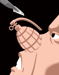
For plastic surgeons hoping to avoid the wrath of a disgruntled patient, the best defense is a good offense, says Richard Goode, MD, professor of otolaryngology at the School of Medicine and chief of otolaryngology at the Veterans Affairs Palo Alto Health Care System. Goode is the author of the article, “The unhappy patient following facial plastic surgery: What to do?” which appeared in the May issue of the journal Facial Plastic Surgery Clinics of North America.
The best way to deal with unhappy patients following facial plastic surgery, Goode advises his colleagues, is to have never operated on them in the first place.
“There are some patients you just want to avoid,” says Goode, who has garnered a certain amount of expertise on the topic through 40 years of occasionally painful encounters with patients. “If you can pick them out before surgery, that’s the best.”
Doctors take seriously the research that shows a small percentage of plastic surgery patients are at risk of experiencing psychological problems after undergoing elective plastic surgery. For some, it leads to depression and occasionally even murder.
“Patients can get very, very angry,” Goode says. “They call you repeatedly, write you harassing letters, leave notes in your mailbox.” In fact, at least a handful of surgeons have been killed by unhappy patients.
Goode says he hasn’t received any death threats, but he has dealt with patients who were extremely angry after surgery even when he was quite happy with the postoperative results.
One woman was so furious after Goode operated on her nose that she visited five other doctors looking for confirmation that his work was shoddy. All five told her the nose work looked fine.
“She kept saying, ‘People stop me in the street. People are laughing at me. My friends all asked me what happened to my nose.’ I tried to get her to see a psychiatrist. That made her more angry,” Goode says.
The key to avoiding the patient with unrealistic expectations, or those obsessing over imagined physical defects, is to use the initial interview as a screening process, Goode says. Take your time, listen carefully and, if necessary, schedule a second appointment.
He also recommends using electronic computer imaging during the interview to help judge the prospective patient’s expectations. And it’s important for every surgeon to have a policy on who pays for revisions when necessary and to make sure this is clear preoperatively, Goode says. —Tracie White
Microbial champion
“Again with the germs, Stanley?” As a young researcher, it was a constant refrain in Stanley Falkow’s life. It’s not that his mother wasn’t supportive. But in the early ‘50s she was skeptical of her son’s choice of a heretofore unknown career in bacterial pathogenesis.

More than 50 years later, there’s a chance she might come around. After all, Falkow, PhD, the Robert W. and Vivian K. Cahill Professor in Cancer Research at the School of Medicine, was just awarded the 2008 Lasker-Koshland Award for Special Achievement in Medical Science.
Sometimes referred to as America’s Nobels, the Lasker Awards are this country’s most distinguished honor for researchers in basic and clinical medical sciences. The Special Achievement award is given only once every two years, to commemorate a life of scientific contribution and service.
“There’s an irreverent, playful joyfulness to the way Stanley does science,” says David Relman, MD, a Stanford professor of infectious disease and of microbiology and immunology, who was a postdoctoral scholar in Falkow’s lab in the late ’80s. “Everyone who meets him feels like they have a personal connection.”
Falkow’s fascination with his chosen field began at around age 11 when he read Paul de Kruif’s Microbe Hunters, which tells of the earliest discoveries of micro-organisms. After reading the book, he was hooked. He arranged a deal with a nearby toy store to work in exchange for a small microscope, and promptly became a member of what was then a relatively small group of bacterial paparazzi.
Since then he’s devoted his life to understanding the microbes’ point of view. He’s particularly fond of promoting the idea that many of the adverse effects of microbial infection are the fault of the host as much as the bacteria. “Disease is a distraction that keeps us from understanding the biology of the relationship between the two organisms,” says Falkow, who is also a professor of microbiology and immunology. “I never met a microbe I didn’t like.”
The inveterate fisherman’s enthusiasm is hard to resist.
“He’s given more to science than just stellar ideas and new insights,” says Relman. “His legacy includes a community of over 100 trainees who view each other as family and who have learned a unique way of looking at the world and of doing science.”
Falkow is most well-known for his work on extrachromosomal elements called plasmids and their role in antibiotic resistance and pathogenicity in humans and animals. But he’s also argued against antibiotics in animal feed, and scientific censorship in the name of national security. When he came to Stanford in 1981, he set aside his study of plasmids to concentrate full time on how organisms as diverse as cholera, plague and whooping cough cause disease in humans.
Falkow’s unassuming nature might have been shaped in part by his mother, now 98 years old. When informed of her son’s Lasker Award she responded, as she has in the past, “Well, Stanley, better you than some stranger I don’t know.” —Krista Conger
Cancer’s dimmer switch
Cancer starts when key cellular signals run amok, driving uncontrolled cell growth. But scientists at the School of Medicine report that lowering levels of one cancer signal under a specific threshold reverses this process in mice, returning tumor cells to their normal, healthy state. The finding could help target cancer chemotherapy to tumors while minimizing side effects on the body’s healthy cells.
Christoph Niemann
The researchers identified a precise threshold level of the signaling molecule Myc that determined the fate of tumor cells in a cancer of the immune system in mice. Above the threshold, high levels of Myc drove immune cells to grow too large and multiply uncontrollably. When the researchers lowered Myc levels below the threshold, the same cells shrank to normal size, stopped multiplying excessively and began dying normally.
A middle way
“This is a new concept,” says Catherine Shachaf, PhD, an instructor in microbiology and immunology who shared lead authorship of the study with colleague Andrew Gentles, PhD, a research associate in radiology. The findings were published in the July 1 issue of Cancer Research.
Identifying the threshold was important because Myc functions in both healthy and cancerous cells as a transcription factor, a protein signal that binds DNA to turn genes on or off. Excess Myc contributes to about 50 percent of human cancers, including malignancies of the immune system and lung.
But Myc is essential, at lower levels, for normal cell function. So, switching Myc all the way off is not an option for treating cancer.
Using genetically engineered mice, the researchers slowly lowered Myc from an elevated, cancer-causing level to the precise point at which tumor cells returned to normal. Near the threshold, they examined many aspects of cell metabolism to obtain a detailed picture of how the cancer cells changed as Myc dropped.
“At the Myc threshold, there is a big change: Programmed cell death becomes dominant over growth,” says Gentles.
“We were able to experimentally prove that we can turn Myc off a little bit, or for a little time, and that’s enough to have a profound effect on cancer,” says co-senior author Dean Felsher, MD, PhD, associate professor of oncology and of pathology. — Erin Digitale
The research was supported by grants from the National Cancer Institute, the National Cancer Institute Integrative Cancer Biology Program, the Leukemia and Lymphoma Society, the Damon Runyon Foundation, the Burroughs Wellcome Fund, a Weiland Family Fellowship and a Flight Attendant Medical Research Institute Young Clinical Scientist Award.
Breath-making development
Five hundred years after Leonardo da Vinci’s intricate drawings of the lung drew the attention of biologists, mathematicians and engineers, researchers at the School of Medicine have published the first description of how the intricate tree of airway tubes develops.
Christoph Niemann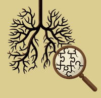
The painstaking work, published in the May 8 online issue of Nature, reveals just three branching patterns that account for the treelike structure of the lung’s airways. Although researchers conducted the work in mice, a better understanding of how lungs develop in mammals could help treat or prevent lung diseases in humans. Knowing the rules that govern how the branching structures form is also essential to researchers’ efforts to regenerate branching tissues, such as lung, pancreas or kidney.
“We believe that setting the airway pattern is essential to patterning all the other tissues in the lung,” says lead author Ross Metzger, MD, PhD. Metzger, now at UC-San Francisco, did the work as a PhD student working with senior author Mark Krasnow, MD, PhD, professor and chair of biochemistry at Stanford.
Figuring out how the branches arise involved long hours staring at individual mouse lungs frozen in development, searching for newly forming buds and analyzing where they were positioned in relation to the main branches. All told, Metzger documented 5,051 newly forming buds, which grew into remarkably similar branching patterns in each lung.
Seeing things
Metzger noticed some consistent patterns. In one pattern, which he calls domain branching, the new branches shot out like bristles on a kitchen bottle brush as the main tube grew longer. Those bristles emerged in four neat rows at right angles to each other, forming a “+” in cross section.
In the orthogonal bifurcation pattern, the tip of a growing branch splits into left and right branches, then each of those split again into top and bottom branches. In cross section, the tips of those resulting four tubes form the corners of a square.
In the final pattern, which he calls planar bifurcation, the initial growing tip divides into left and right branches, then each divides again into a left and a right branch. The four resulting branches lay on the same plane as if they were drawn on a piece of paper.
The emergence of three consistent patterns is a surprise in the field, Metzer says. “The bias in the field has been that the pattern is random or just a matter of one simple branching mechanism. Somehow people weren’t thinking about generating patterns,” he says. —Amy Adams
The work was funded by grants from the National Institutes of Health.
Dr. Doctor
Oscar Abilez, MD, wants to look back on the days of surgeries with inorganic stents and valves as a quaint era.
“Titanium, carbon-based material, Teflon, silicone — everything has been inert,” he muses. “But over the last 10 years we’ve transitioned from nonliving to living replacement parts.”
During his two years as a resident in general surgery at Stanford Hospital, Abilez assisted in thoracic and vascular surgeries, and he knows how dying heart muscle and blocked arteries look and feel.
Now, after putting the remaining three years of his residency on hold to earn a PhD in bioengineering, Abilez is trying to grow tissue that could be used to heal and renew the body’s critical pump and its blood supply. “On any given day, we can take undifferentiated embryonic stem cells and start to process heart-muscle and blood vessel cells,” he says. “You can look into the culture dish and nothing’s beating, but when you come back the next day, it’s beating on its own — it’s fascinating.”
Cross-training trainees
Abilez just completed his first year of Advanced Residency Training at Stanford, a program launched in the fall 2007 that helps residents and clinical fellows earn PhDs. ARTS’ goal is to break down the growing barriers between clinical and basic research — a trend noted by the National Institutes of Health in its Roadmap for Medical Research. “It’s about how we can bridge the work of laboratories on campus with what goes on in the hospital,” says ARTS director Sam Gambhir, MD, PhD, professor of radiology.
At an annual cost of some $120,000 per fellow for the five years it can take to earn a PhD, ARTS could be an expensive experiment. But Gambhir says School of Medicine Dean Philip Pizzo, MD, has agreed to have the medical school underwrite the cost as an investment in “improving our entire educational process.”
Abilez is one of the first two ARTS fellows. Two more are set to begin this fall and more next year as word of the program circulates. Current residents or clinical fellows at Stanford are eligible.
Unlike students enrolled in traditional MD/PhD programs, who do their doctoral work while they’re still in medical school, ARTS fellows complete several years of clinical work before they start their research. “You’ve seen something at the bedside that doesn’t make sense, and you can say, ‘Let’s look at it in the lab,’” says ARTS fellow Christine Ham, MD. “‘Let’s see, molecularly, what it means.’”
Gambhir says Abilez and Ham are just what the doctors at the school and at the hospital ordered. “They have a commitment to medicine, and they bring to the lab environment a perspective on clinical issues and problems,” he says about the two future physician-scientists. More important, “they really want to do research.” — Diane Rogers
Tear repair
Until now, U.S. doctors relied largely on hunches to choose the best treatment for women whose vaginal tissues were torn severely during childbirth — a fairly common occurrence. But a recent medical center study offers hard data: A single dose of antibiotics can significantly aid healing.
As many as one in five women suffer severe vaginal tears while giving birth. In the study, those who received the antibiotic endured roughly one-third as many infections or other wound-healing complications two weeks after surgical repair of their tears.
“If you add an infection, or a breakdown of the surrounding tissues, it’s a huge burden on the emotional and physical well-being of a new mother.”
“Recovery from these tears can be painful and problematic,” says senior author Yasser El-Sayed, MD, associate professor of obstetrics and gynecology and associate chief of maternal-fetal medicine at Lucile Packard Children’s Hospital. “If you add an infection, or a breakdown of the surrounding tissues, it’s a huge burden on the emotional and physical well-being of a new mother.”
The study was published in the June issue of Obstetrics and Gynecology.
Vaginal tears, which occur between the vagina and the anus, are classified in severity according to their length. Third-degree tears extend into the muscle of the anal sphincter and fourth-degree tears reach the rectum. They are surgically repaired immediately after delivery.
El-Sayed and his colleagues conducted a randomized, double-blind study in 147 women who experienced third- or fourth-degree tears while delivering infants at either Packard Children’s Hospital or Santa Clara Valley Medical Center. After agreeing to participate in the study, the women were randomly assigned to receive a one-time intravenous infusion of either antibiotic or placebo during the repair of their tear.
The researchers found that four of 49 patients (8.2 percent) treated with antibiotics and 14 of 58 patients (24.1 percent) who received the placebo showed symptoms of infection or breakdown two weeks after the repair. The remaining 40 women did not return for their scheduled follow-up appointments, but the difference between the two returning groups was statistically significant.
“We’re excited because it’s such a simple intervention,” says El-Sayed.
Although the study was relatively small, the results detected an important difference in outcome. Until now, physicians were divided as to whether antibiotic treatment was helpful for these women, and most health-care providers made their own choices. —Krista Conger
The research was supported by Stanford’s Department of Obstetrics and Gynecology and the Santa Clara Valley Medical Center.