 |
|||
| Stanford Medicine |
| Current Issue |
| Contact Us |
| ALL Issues |
| 2008 Spring |
| 2007 Fall |
| 2007 Summer |
| 2007 Spring |
| 2006 Fall |
| 2006 Summer |
| 2006 Spring |
| 2005 Fall |
| 2005 Summer |
| 2005 Winter |
| 2004 Fall |
| 2004 Spring |
| 2003 Fall |
| 2003 Summer |
| 2003 Winter |
| 2002 Fall |
| 2002 Summer |
| 2002 Winter |
| 2001 Fall |
| 2001 Winter/Spring |
| 2000 Fall |
| 2000 Spring |
| 1999/2000 Winter |
| 1999 Summer |
| 1999 Spring |
A quick look at the latest developments from Stanford University Medical Center
![]() The new fat farm
The new fat farm
![]() Glimpse a muscle's inner flex
Glimpse a muscle's inner flex
![]() An anthrax lesson
An anthrax lesson
![]() Master's of science — in medicine
Master's of science — in medicine
![]() The beat goes on
The beat goes on
![]() Stanford med gets an A+
Stanford med gets an A+
The new fat farm
In the middle of John Steinbeck country, the “salad bowl of America,” most of the Mexican farmworkers who harvest the fruits and vegetables that feed the nation aren’t eating enough of it themselves.
Paul Blow |
|
 |
|
Underpaid and over-worked, Salinas farmworkers are eating at fast-food restaurants where the food is high-fat but low-cost. As a result, the Latino farmworkers — particularly those single, young men living in the agricultural labor camps — are facing a very American problem: obesity.
“They often eat someplace that’s cheap and fast with high fat content,” says Marilyn Winkleby, PhD, associate professor of medicine at the Stanford Prevention Research Center, who has spent years visiting farm laborers in the Salinas Valley. “Their jobs are becoming increasingly mechanized and less active.”
Winkleby is the senior author of a study published in the February issue of Ethnicity and Health examining the changes that occurred between 1990 and 2000 in cancer-related health behaviors within the Salinas Latino population, most of whom are of Mexican origin. The study surveyed almost 2,000 Latino women and men from both the community at large and the Latino population within 29 agricultural labor camps.
The goal was to detect changes in diet, physical activity, smoking and alcohol use as well as cancer health screenings in order to help design future public health interventions. Latinos, the country’s fastest-growing minority group, suffer disproportionately from poor health exacerbated by poverty, poor education and a lack of health insurance and medical care, according to the study.
Not all the news was bad. Among the positive results were continued low rates of smoking among women and men and a substantial drop in alcohol consumption among men. There was also a shift away from whole-fat to lower-fat milk, and a drop in the use of meat fat for cooking. There were also significant increases in screenings for both breast and cervical cancer.
But most striking among the results were the obesity rates. By 2000, 70 percent of the men in the community sample were overweight or obese, as were 64 percent of the women in the community sample and 60 percent of the men in labor camps. Within the camps, there was an increase in obesity of 90 percent.
“If you want to effect change it’s not enough to educate people,” Winkleby says.
“Individual behavior is influenced by the environments in which you live. Men living in the labor camps are primarily single, poor; they live in housing without cooking facilities.” — TRACIE WHITE
| ![]() Back
to Top |
Back
to Top |
Glimpse a muscle's inner flex
Dahlia Weiss has invested years in her thesis project, which involves creating a dynamic model of the motor protein known as myosin. But she already knows that when she gets her PhD, the project won’t be anywhere close to completion.
Ethan Hill |
|
 |
|
“For me, it would be great to build a model that has the same biophysical properties as myosin,” Weiss says. “Beyond that, I’ll leave the rest to someone else.”
Weiss is part of a team headed by Michael Levitt, PhD, professor of structural biology, that’s studying myosin, which drives a number of critical cell processes including muscle contraction and cell division.
Of the 13 known types of myosin, Levitt is focusing on myosin 2 or conventional myosin, which reaches out and pulls itself forward along filaments of actin, a major protein component of the cell cytoskeleton.
Myosin 2 molecules cause the contraction of skeletal muscle by working together in chains, which simultaneously pull themselves along actin tracks from opposite ends of each muscle fiber. By representing the behavior of a single molecule, the model will offer an up-close view of the force-generating agent underlying all motor movement.
Scientists have known the three-dimensional architecture of myosin’s 10,000-plus atoms since the 1990s, and they have also known the precise structures of myosin 2 at the beginning and end of its pulling action. Levitt’s team is using computers to help fill in the motion between these pictures.
“If you have certain atoms sitting in certain locations and if you know something about the forces between the atoms, then you can create computer-based simulations that predict how everything will interact and move,” says Levitt.
Programming these simulations is anything but straightforward. “You need a lot of biological background and intuition to know if what you are doing makes any sense at all or if it is computer-generated junk,” says Weiss.
Still in the early stages of the project, researchers are grappling with one of the key challenges of model building: defining an appropriate level of complexity. So far the team has reduced the molecule’s 10,000 atoms to 60 rigid groups of atoms, which they would ultimately like to distill to five or six groups.
Once researchers have honed the contours of their model, they’ll begin fine-tuning the movie of myosin’s action by testing how well the model can mimic known experimental findings. Ultimately, though, Levitt would like the finished model to be able to make predictions that help inform experiments at the bench. — PAM LOWNEY
The research is supported by the National Institutes of Health through the Simbios National Center for Biomedical Computing at Stanford.
| ![]() Back
to Top |
Back
to Top |
An anthrax lesson
When spores sent through the mail in 2001 caused 11 people to contract anthrax — ultimately killing five of them — the death rate was well below the historical mortality rate, which approached 100 percent. Many assumed that access to modern intensive care units and powerful antibiotics made the difference.
Paul Blow |
|
 |
|
But a review by researchers at the Veterans Affairs Palo Alto Health Care System and the School of Medicine pinpointed the true reason: rapid diagnosis and initiation of antibiotic treatment within the first few days of anthrax symptoms.
The researchers found that once anthrax progresses to its advanced stage, patients are almost certain to die from it, even if they receive the best care modern medicine has to offer. They also found that drainage of fluid from around the lungs is a key procedure associated with anthrax patients’ survival.
The study, published in the Feb. 21 issue of the Annals of Internal Medicine, underscores the importance of detecting anthrax early, educating medical personnel about its symptoms and treatment, and ensuring efficient distribution systems that can deliver antibiotics to patients within hours of a bioterrorist attack.
“Even with our modern intensive care, once you’ve reached the advanced stage of this disease, you’re probably going to die. That’s why it’s crucial to start antibiotics within the first few days,” says lead author Jon-Erik Holty, MD, a fellow in pulmonary and critical-care medicine at Stanford, who did the research during a fellowship at the VA-Palo Alto.
Anthrax is an acute infectious disease caused by the spore-forming bacterium Bacillus anthracis. It progresses in two phases: an initial phase lasting about four days, which produces flu-like symptoms including cough, fever and chills; and an advanced phase, which causes respiratory distress and shock.
For the study, the researchers sought out all published reports of inhalational anthrax from 1900 to 2005; they ended up with 82 confirmed cases from 15 countries. For each case, the researchers documented the year; the patient’s age, sex and nationality; the presenting symptoms and the stage at which anthrax was diagnosed; the type of treatment given and the timing of treatment; and whether the patient survived.
The overall death rate was 85 percent, but for patients who progressed to the advanced phase, the death rate was 97 percent, even among patients who received supportive care in a hospital intensive care unit.
Timely antibiotic treatment was the key to patients’ survival. When antibiotics were begun within two days of initial symptoms, approximately 20 percent of patients died. When treatment was begun at four days, mortality was about 58 percent, and at six days it was nearly 80 percent. Among all the anthrax patients who survived, 80 percent had fluid drained from around their lungs. — SARA SELIS
| ![]() Back
to Top |
Back
to Top |
Master's of science — in medicine
If translational medicine is going to live up to its promise, it needs more people to do the translating — scientists who really know the language of the clinic and the lab.
Stanford’s new master’s of science in medicine program aims to help fill that gap by training a new generation of translational scientists: researchers who will turn basic discoveries into treatments for patients. The program, one of the few of its kind, was approved in December by the university’s faculty senate.
The program will allow students on a PhD track in the basic sciences to complete rigorous pre-clinical course work in human biology and then spend one to two intensive months doing clinical rotations.
“We have all these basic biomedical advances, but we’re not successful in converting them into treatments for patients.
I think a huge part of the problem is we haven’t taught researchers about basic human diseases,” says Ben Barres, MD, PhD, a professor of neurobiology and of developmental biology, who initiated the program and serves as its director.
Barres says graduate students often come in feeling passionate about human disease but channel their energy into bench work, never getting a chance to learn about human biology or the problems that afflict people. Many have asked for a program that gives them more of the human side of science, he says.
The master’s program would add a year to 18 months to the usual four or five years that PhD students spend at Stanford, but would still be significantly shorter than a traditional MD/PhD program.
The first crop of students will enter the program this fall. Six students will be accepted the first year and up to 10 students per year after that, all selected from applicants to Stanford’s 12 PhD programs in biosciences, bioengineering, chemistry, physics and biomedical informatics.
The Stanford program, which will cost $74,000 per year per student, will be funded largely through private grants. The Howard Hughes Medical Institute has awarded $750,000 over four years to the degree program, and Amgen Foundation has agreed to underwrite the costs for three students. The medical school also has made a financial commitment to the program.
More information on the program is available on the Web at http://msm.stanford.edu/.
— RUTHANN RICHTER
| ![]() Back
to Top |
Back
to Top |
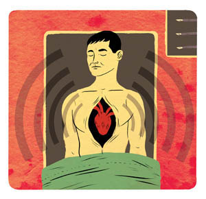
The beat goes on
Heart surgeon Kai Ihnken reaches into the chest of a 78-year-old man undergoing triple bypass surgery and lifts the beating heart, then cranes his neck to the side, searching for the right spot on the back of the heart to attach the next vessel.
Paul Blow |
|
 |
|
Ihnken, MD, clinical assistant professor of cardiothoracic surgery, uses an innovative technique called “beating-heart” surgery for coronary bypass. It replaces the more conventional use of a heart-lung machine, which allows surgeons to stop the heart during surgery.
“It’s very, very technically demanding,” says Ihnken. “Surgeons don’t want to put up with the stress. But it’s so beneficial for the patient.”
Beating-heart surgery has garnered renewed attention recently as the trend toward less invasive methods of heart surgery grows stronger.
“We need to be researching this,” says Robert Robbins, MD, chair of the cardiothoracic surgery department, who hired Ihnken to investigate the potential benefits of beating-heart and robotic surgery. “We need to make it available to our patients.”
Most heart surgeons put a patient on a heart-lung machine, aka “the pump,” for bypass surgeries. The pump allows the surgeon to stop the heart by keeping blood circulating throughout the body while the doctor sews on new vessels. It’s easier for surgeons to perform because the heart is not moving.
In beating-heart surgery — currently used in about 22 percent of U.S. bypass procedures — the surgeon uses a stabilizer (a small device with a suction cup attached) to minimize the motion of the heart where the stitching is done.
Proponents of beating-heart surgery point to the risks associated with conventional bypass surgery, such as blood loss, stroke and kidney failure. Because the body doesn’t have the added stress of being put on a heart-lung machine, beating-heart surgery can reduce these risks, especially among high-risk groups which include the elderly and, surprisingly, women, Ihnken says.
“Women have double the mortality rates following bypass,” he says.
A recent report in the journal Innovations found that the beating-heart technique is as safe as conventional heart surgery, with fewer complications and lower costs. Still, scientific evidence is not yet conclusive, says Robbins, who wants to see more research done before he’s convinced that beating-heart surgery beats out the heart-lung machine.
“It’s just so difficult,” says Robbins, who used the procedure when it was first developed a decade ago, but now uses it only for selected cases. “I don’t have the time to devote to it. You need to do it often to be good at it.”
Which is exactly what Ihnken plans to do. He’s completed 130 off-pump surgeries, including all of his bypass work. “I really want to build Stanford as an off-pump center,” he says. — TRACIE WHITE
| ![]() Back
to Top |
Back
to Top |
Stanford med gets an A+
After 18 months of preparation, the School of Medicine passed its big test with high marks, earning full accreditation for the next eight years.
The news arrived in mid-March from the Liaison Committee on Medical Education, the national group that accredits medical schools in the United States and Canada. The LCME issued a 205-page report detailing the school’s strengths, as well as noting a few areas where it hopes to see progress.
“This was an outstanding report,” says Oscar Salvatierra, MD, professor of surgery and of pediatrics (emeritus) and the effort’s faculty leader, noting that it was an “incredible turnaround from 1997,” when the LCME cited a number of significant problem areas.
The success, he added, can be credited in part to the “great faculty and students who came together to present the best the medical school had.”
The school began preparing two years ago for the review, which involved more than 200 faculty, staff and students. Some 3,000 pages of documentation and a 50-page report were submitted before the LCME team’s site visit in October 2005.
The final report praised Dean Philip Pizzo, MD, for his commitment to medical education and for serving as a catalyst for curriculum reform. It noted that he has committed significant financial resources to education, encouraging faculty teaching through financial support to departments in return for faculty time in the classroom. It also lauded the many interdisciplinary faculty collaborations.
Among other strengths, the report cited the school’s investment in information technology and the creation of a “library without walls,” giving students and faculty access to information anytime, anywhere. The new information technologies help faculty use various computer-based applications to teach and evaluate students, it noted. The LCME also applauded the school’s effort to minimize student debt. Average medical student debt at Stanford is less than half that of other private schools.
The LCME asked for a progress report in 2007 addressing several areas, including resolving scheduling conflicts between students’ regular classes and work in their scholarly concentrations, as well as an update on the new system for evaluating students’ clinical skills.
Cited repeatedly in the past for inadequate facilities, the school was finally commended for “significant improvement,” including plans for the 120,000-square-foot education building, which were approved in concept by the university’s trustees in October. The LCME also lauded the $17 million investment in renovations to classrooms, the Fleischmann laboratories and anatomy labs. — RUTHANN RICHTER
| ![]() Back
to Top |
Back
to Top |
Comments? Contact Stanford Medicine at