Alumni profile: William Northway
One man's extraordinary career in children's film – X-ray film, that
is
|
Leslie Williamson |
|
 |
 |
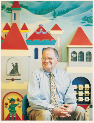
William
Northway’s magic kingdom — the radiology department waiting
room at Lucile Packard Children’s Hospital. |
By KRISTA CONGER
It was a medical mystery. On the X-ray, the infant’s lungs looked like a cross-section of a kitchen sponge: pockmarked with holes cobbled together with a spider web of thin filaments. The year was 1964, and pediatric radiology instructor William H. Northway Jr., MD, class of ’57, had never seen the distinctive pattern before. • “Do you mind if I investigate this a bit?” Northway asked neonatologist Philip Sunshine, MD, who had brought him the film. And thus a career was born.
Northway’s subsequent discoveries would change the way premature babies with lung disease are treated worldwide. His ability to weave research with patient care and teaching was honored when he received the Stanford Medical Center Alumni Association’s J.E. Wallace Sterling Alumni Lifetime Achievement Award in May.
It’s no great surprise that Northway, who served as the director of pediatric radiology at Lucile Packard Children’s Hospital from 1994 to 1998, remains associated with the medical school. The Northway family is something of a Stanford institution. Northway’s father, uncle and a brother all received bachelor and medical degrees from Stanford, his mother was a nurse at Stanford after graduating from the university’s former nursing school, and another brother graduated from Stanford’s architecture program. His father was a chairman of physical medicine at Stanford and physician for several of the school’s athletic teams. And Northway’s own two children graduated from Stanford.
Northway’s interest in radiology began when, as a Stanford undergraduate, he abandoned his original plan to be a mathematician. “It quickly became clear that, although it was fun to think about, there were a lot of other people who were better at it than I,” says Northway, who decided to go to medical school instead. A senior year course on radiotherapy treatment of cancer with the new chair of radiology, Henry Kaplan, MD, set him firmly on course.
“Henry was an extremely bright guy, and he made radiology a very exciting field,” says Northway. “He would assign six or eight scientific articles to read with differing opinions on the same topic, and he’d go through them with us: this one didn’t have controls, this one dealt with only low-grade malignancies, etc. It was a wonderful learning experience.”
Kaplan’s influence propelled Northway first into medical school and then a residency in radiology at Stanford, during which the medical school relocated from San Francisco to Palo Alto. After a stint as a diagnostic radiologist in the Air Force at Biloxi, Miss., Northway decided in 1963 to home in on pediatric radiology.
When Sunshine approached Northway in 1964, Northway had just returned from a two-year pediatric radiology residency at Paris’ Hospital des Enfants Maladies to replace Stanford’s first pediatric radiologist, Robert Evans, MD. Even with all his experience, though, Northway hadn’t seen lungs like these. He was further perplexed when Sunshine mentioned that this spongelike pattern was becoming more common in premature babies three to four weeks after birth.
Ventilation for babies
That Sunshine’s small patient was alive at all was a dramatic testimony to the success of a new technique for treating premature babies that was gaining popularity around the country. About a year earlier the same tiny baby would have been placed in an oxygen tent and given fluids and antibiotics. This treatment would save a few lives, but many more would die when the tiny air sacs in their immature lungs collapsed. The subsequent spiral of airway cell damage and death left behind transparent, membranelike deposits that led this type of respiratory failure to be called hyaline membrane disease.
In 1963 physicians hit upon the idea of using a mechanical ventilator developed for use in adults to keep the airways of premature babies open long enough for the lungs to mature and function on their own.
“At the time, we were in the dark as far as how to ventilate babies,” says Sunshine. “It was all trial and error.” Although they had to guess at the appropriate pressure and oxygen concentrations for the infants, more of what were then considered to be very small preemies (about 5.5 pounds) were surviving the first harrowing days after birth than in the pre-ventilator days.
Then came the puzzling X-rays.
“Textbooks at the time taught that hyaline membrane disease did one of two things: the babies either died within three or four days of birth, or they got better and their lung X-rays appeared normal after a few weeks,” says Northway. “I kept wondering ‘Why is this so different?’ ”
Northway eventually hit upon a possible explanation: all the babies with the spongelike X-rays had been on a ventilator receiving at least 80 percent oxygen for seven days or more. Those who received fewer than seven days of treatment had normal lung X-rays.
“When the data fell out like that, it was like a light bulb going on,” says Northway, who termed the new chronic lung condition bronchopulmonary dysplasia. The diagnostic radiology department’s unusual policy of allowing faculty members to devote two days per week to research allowed him to duplicate the effect in newborn mice and guinea pigs, showing that high concentrations of oxygen actually slowed the growth and development of the lungs by inhibiting DNA synthesis. The 1969 paper describing the results in guinea pigs was named a radiology classic by readers of Investigative Radiology.
Quick change
Although the findings were at first controversial, change came quickly. “People lowered the oxygen concentration and reduced the ventilation pressure,” says Northway, who at the time was an assistant professor of diagnostic radiology under Herbert Abrams, MD. “They developed devices to easily monitor oxygen concentrations in the blood, and modified the adult ventilators to be more suitable for tiny babies.” As a result, the prevalence of bronchopulmonary dysplasia began to decline and now occurs only in the most severely ill, very premature infants.
Years of research by Northway, who would go on to serve as director of diagnostic radiology and associate chair of the radiology department from 1976 to 1980, and his colleagues at Stanford have proven that although most infants with the condition grow up to have seemingly normal lung function, some have persistent pulmonary problems including wheezing and shortness of breath when exercising. They can also have permanent airway thickening and narrowing. The long-term effect of this dysfunction is not known; the oldest of the affected children are only in their late 30s or early 40s.
“Of course we warn all the kids in the study and let them know
they probably shouldn’t ever start to smoke,” says Northway,
with his trademark concern for his patients.
“Bill is one of the few radiologists who would examine a patient when they came down for a procedure, and he took great care to keep the tiny babies warm and comfortable,” says Sunshine. “He’d pick up things that the pediatricians missed. One time there was a little kid with nodes along one of his nerves. We couldn’t figure out what was wrong with him. When Bill told us he had neurofibromatosis, some of the other physicians said, ‘What are you doing examining our patient?’ But it turns out he was right.”
Examining kids takes a special kind of doctor. “You can’t say to an infant, ‘Stand up now, OK, hold your breath, I’m going to take a picture,’ ” says Northway. “I like working with kids, but not everybody enjoys it. It’s not a piece of cake.”
When Northway retired from Stanford in 1998, he headed to the Pacific Northwest for a fly-fishing vacation. But he soon found himself back at Stanford. When he heard his department was short-staffed he agreed to teach residents and read films one to three mornings a week.
“Working with other people who enjoy working with children is very rewarding,” he says. “They are all very committed to the kid in the family.”
As is Northway.
Comments? Contact Stanford Medicine at
 Back
To Contents
Back
To Contents