Brain Flash
So that’s how nerve cells transmit information so quickly
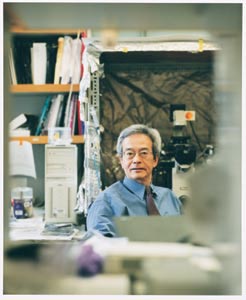
By Mitzi Baker
Photographs by Leslie Williamson
Kiss-and-run. While the term brings to mind spouses being dropped off
at a commuter rail station, neuroscientist Richard Tsien, PhD, uses it
to explain a newly established method that neurons use to relay information.
Tsien’s new finding overturns long-held beliefs about how the nervous
system works and helps explain how neurons in the brain can transmit
information with lightning speed.
Neurons communicate with each other by releasing chemicals across the
junctures that separate one nerve cell from another – the synapses. “The
synapse is one of the underexplored frontiers of neuroscience,” says
Tsien. While scientists know that individual neurons release chemicals – known
as neurotransmitters – to communicate to others across the synapse
gap, not a lot is known about how the nerves move these neurotransmitters
from inside the cell out into the synapse.
Until now, textbooks explained information transfer across a synapse
this way: Inside a neuron, membrane-enclosed bubbles carrying information-relaying
neurotransmitters fuse with the neuron’s surface membrane and expel
their contents into the synaptic cleft, the narrow space between transmitting
and receiving nerve cells. The bubbles, known as vesicles, can then pinch
off again and reform – a process that takes at least a few minutes.
The problem with this theory is that it fails to account for the high
rate of information transfer in the brain, where the numerous tiny neurons
might have only 30 active vesicles each.
“We followed a different hypothesis,” says Tsien, the George
D. Smith professor of molecular and genetic medicine. Tsien and his team
explored an unproven mechanism that neuroscientists recognized but didn’t
take seriously. “It was heretical enough that the vast majority
of neuroscientists didn’t believe it.”
Tsien’s theory, known as “kiss-and-run,” envisions
no collapse of the vesicle at all; instead, a vesicle opens briefly to
let some neurotransmitter out and then closes and can quickly be readied
to release more neurotransmitter soon thereafter. Tsien, along with Alexander
Aravanis and Jason Pyle, students earning MD/PhDs in the Stanford Medical
Student Training Program, have provided the first evidence of this type
of release. Their findings were published in the June 5, 2003, issue
of Nature.
Neurobiologists have traditionally used the retina of fish for their
studies of neurons because these neurons are big and easy to handle.
Much of what we know about how synapses work comes from these specialized
fish neurons, which can contain a million vesicles each. But the environment
in the fish retina is quite different from that found in the human brain,
where neurons are compact and abundant, says Tsien. His team used rat
brain neurons, cells more similar to those found in the human brain,
to observe transmission across the synapses.
The traditional method of studying synapses relies on monitoring the
cells’ electrical signals, a method that is cumbersome and indirect. “We
decided to take the bull by the horns and study brain synapses directly,” Tsien
says.
So his group did something that hadn’t been done before, which
is to study the vesicles individually and watch them release their contents
in real time. They devised a method that trapped fluorescent dye within
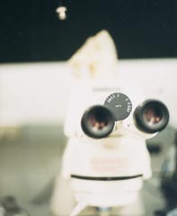
a vesicle so they could track that vesicle’s life cycle. While
viewing the vesicle under the microscope, the researchers directly measured
the decrease in fluorescence as the vesicle released the dye.
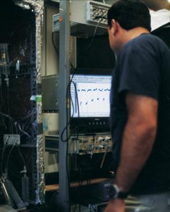
Top-notch equipment was key to the group’s success. Medical students
Pyle and Aravanis, both of whom have studied engineering, used their
skills to fine-tune the microscope and adjust the camera and data-acquisition
processes to work efficiently. “They cleaned up the optical system
so that the signal-to-noise ratio was really amazingly good. That’s
what allowed the signal to be detected,” says Tsien.
“There is a strong component of physics and engineering meets
biology here,” he adds. Although no one has been able to track
a vesicle’s fate in real time before, Tsien credits Stephen J.
Smith, PhD, professor of molecular and cellular physiology, for laying
the groundwork to study single vesicle dynamics using fluorescent dye.
The trio’s findings provide the evidence that the proposed kiss-and-run
mechanism really does work in neurons taken from the brain; that a vesicle
can open up, recover within a second or two and then fire again. The
team also found support for the older hypothesis for vesicle transmission.
Nerves seem to use both methods.
“That re-use, that ability of a vesicle to open up a second or
third time is what we think makes vesicles much, much more efficient
than previously thought at releasing neurotransmitter over and over again,” says
Tsien.
Tsien says that there remain many challenging questions to answer. For
now, he says, their results go a considerable way toward explaining how
tiny synapses in the brain can transmit a vast amount of information
very quickly. “I wouldn’t say that every mystery is completely
cleared up, but it certainly helps that vesicles can be re-used,” says
Tsien. “I don’t want to claim that this is the end of the
story; it’s really the beginning.”
Comments? Contact Stanford Medicine at
 Back
To Contents
Back
To Contents