Upfront
Detection without radiation
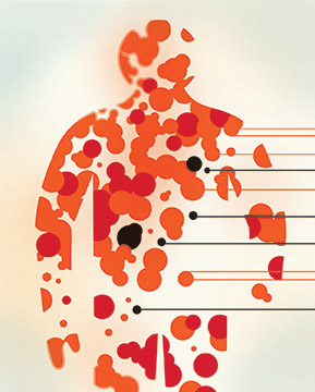
After learning they have cancer, lymphoma patients usually get scans to locate tumors throughout their bodies. But the standard imaging method, whole-body PET-CT, has a big drawback: One scan exposes the patient to as much ionizing radiation as 700 chest X-rays.
This is especially risky for children and teenagers, who are particularly vulnerable to radiation because they are growing. They are also more likely than adults to live long enough afterward to develop a secondary cancer.
That’s why researchers at the School of Medicine and Lucile Packard Children’s Hospital Stanford developed an imaging technique that uses no radiation at all. The method, described in The Lancet Oncology, is a modification of magnetic resonance imaging that employs a novel contrast agent.
The new agent consists of injected nanoparticles of iron, which are already FDA-approved to treat anemia. On MRIs, they cause blood vessels to appear brighter, providing anatomic landmarks. The nanoparticles also cause healthy tissues such as bone marrow, lymph nodes, liver and spleen to appear darker, making tumors stand out.
The nanoparticle-enhanced scans had similar levels of sensitivity, specificity and diagnostic accuracy to whole-body PET-CT. Although more evidence of the technique’s efficacy is needed before it will be adopted, there are no technologic hurdles to its use.
“It’s really exciting that this will soon be clinically applicable,” says radiologist Heike Daldrup-Link, MD, who led the research.