In Brief
Rodin’s virtual hands
Art and medicine meet in augmented reality
Video Extra
High tech scans reveal Rodin's hands
While James Chang, MD, was doing his surgery residency at Stanford, he took to playing a game of sorts with the campus Rodin sculptures: He’d come up with specific medical conditions represented in their hands. He had plenty of hands to choose from as Stanford’s Cantor Arts Center holds one of the world’s largest Auguste Rodin collections, with 200 of his sculptures, including The Thinker and The Gates of Hell.
“I began to notice that most of the hands looked like the conditions I was treating, from fractures to malformations to tumorous growths,” Chang says.
Rodin’s Large Left Hand appeared to have some broken metacarpals. He speculated that Large Clenched Hand, a sculpture frozen in a painfully exaggerated and abnormal posture, had Charcot-Marie-Tooth disease, an inherited neurological disorder. From sculpture to sculpture, hand to hand, the surgeon proceeded to identify a ganglion cyst, a thumb amputation, a stiff joint and other conditions.
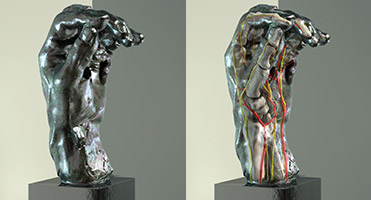
Photographs by Sarah Hegmann
When he became a surgery professor at Stanford, his hobby turned into a teaching tool, which he incorporated into the undergraduate course Surgical Anatomy of the Hand: From Rodin to Reconstruction.
Now Chang’s diagnoses are part of the inspiration behind an exhibit at the art center, Inside Rodin’s Hands: Art, Technology, and Surgery, celebrating the connection between Rodin’s fascination with the human form and medicine’s fascination with human anatomy. The center’s program includes a “virtual operation” to fix the perceived broken fingers on the Large Left Hand and a 3-D, augmented-reality model showing how the statue would look post-surgery. The exhibit runs through Aug. 3.
“Art is informing medicine in the exhibit, and medicine is informing art,” says the museum’s director, Connie Wolf. “It’s unlike anything we’ve ever done before.”
Stanford has a history of scientists using art to inform their teaching and learning, stretching back at least to the early 1990s, when surgery professor Robert Chase, MD, and Rodin authority Albert Elsen, PhD, challenged medical students to find clues of medical conditions in the Rodin sculpture garden — located conveniently near the medical school.
The new exhibit builds upon this history, says Paul Brown, DDS, consulting associate professor of anatomy. Brown, together with exhibit production manager Matt Hasel and medical artist Sarah Hegmann, used CT scans from Stanford hand clinic patients to create virtual sculptures showing the hands’ supposed internal anatomies.
Rodin studied anatomy like other art students of his day, and he spent time at the Musée Dupuytren in Paris (a museum of anatomical items illustrating diseases and malformations), says Rodin expert Bernard Barryte, the exhibit’s curator. So were Rodin’s hand sculptures based on living models or did they grow from his imagination? According to Barryte, no one knows for certain.