
SUMMER 2010 CONTENTS
Home
Transformers
How teaching hospitals could lead medicine’s metamorphosis
No holes barred
Interest grows in using natural openings for surgery
The healing hand
Putting the physical back in the physical exam
Take the tube
Mass transit for lab samples
Code green
New hospitals blend healing and conservation
On the record
The nation’s health information technology leader on the future of patient data

DOWNLOAD PRINTABLE
ISSUE (PDF)

Virtual cell
Christian Northeast
In a purely logistical sense, biology can really get in the way of biological research. Cells must be cultured, nurtured and then perturbed according to an experiment’s protocol. It can be slow, demanding and expensive work.
While most kinds of scientists and engineers can use computer simulations to speed up their research, there has been no such thing as a virtual cell. Now one is taking shape in the research group of Markus Covert, PhD, assistant professor of bioengineering. In a matter of months, he expects to unveil the first “whole-cell” computer model of an organism.
“To me, this is a dream,” says Covert, who got the idea from a New York Times article he read 10 years ago suggesting that the ultimate challenge would be to make a computer model of a cell “because that implies biological understanding at a fundamental level.”
The microbe that Covert’s team is simulating is Mycoplasma genitalium, a sexually transmitted parasite. The nasty bug is a perfect candidate to simulate because of its simplicity. It has only 521 “coding regions,” the segments of DNA with complete instructions for doing things such as making proteins. Most of those, about 420, are understood by biologists. As 2010 began, Covert’s model accounted for more than 300 coding regions with more joining the model every week.
In other words, about 75 percent of what biologists know about Mycoplasma genitalium is now being simulated on Covert’s computer. That’s already enough to give him two things: confidence about integrating the rest of the coding regions, and a simulation that already works well enough for his group to ask biologically interesting questions about the organism, such as why it grows so much more slowly than its close relatives.
Success with this model will lay the groundwork for tackling more complicated and biologically important organisms, such as yeast and E. coli. Ultimately, Covert’s goal is to give biologists the same computational simulation tools that allow peers, such as aircraft designers and nuclear physicists, to perform accurate experiments that are cheaper and quicker (and often safer) than tests with real materials and environments.
“You discover things a lot faster with a computer model than you do without,” he says.
Covert’s model incorporates many different systems that are at play in a cell, and will therefore give researchers a general platform for investigating a much larger set of questions. In fact, one of Covert’s goals is to find out how cells age and why low-calorie intake corresponds to greater health and longevity.
As the Mycoplasma genitalium model comes together, Covert’s group is also working on creating a graphical interface that provides users with a dashboard view of what’s going on in the cell. The collection of graphs and dials and other visual indicators will also give researchers an easy way to tweak parameters, such as removing a coding region to perform experiments.
— David Orenstein
The research is funded by a National Institutes of Health Director’s Pioneer Award.
Unbiased ed
The School of Medicine has developed a new model for continuing education for physicians that aims to ensure that corporate donors exert no influence over the courses. Now the school is putting the model to the test, using a large industry grant to develop a new curriculum for continuing medical education classes.
In September 2008, Stanford became the first medical school in the country to place stringent limits on industry support of its CME programs — the courses doctors take to keep their licenses. Nationwide, health-care companies provide about $1 billion a year to support CME.
Stanford’s new approach prohibits industry from directing its funding to support specific programs. Instead, the school is exploring educational models that involve industry but would be guided by the needs of physician-learners rather than by commercial interests, says Robert Jackler, MD, the school’s associate dean for postgraduate medical education.
“We believe that the education of practicing physicians should be based solely on the best scientific evidence presented in a fair and balanced way,” says Jackler, professor and chair of otolaryngology. “Unfortunately what’s happened is that the partnership with industry has led CME astray, to the point where the curricula are too often biased toward business interests.”
No strings
Under the new model, Stanford will use a new, $3 million, three-year grant from the pharmaceutical company Pfizer to design and implement a curriculum that uses advanced technologies and teaching methods, including simulated and immersive learning tools. The grant comes with no conditions, and the company will not be involved in either selecting topics or shaping the curriculum.
The overall goal of the program is to improve patient care and outcomes, with a focus on specific patient-care issues identified by Stanford physicians, Jackler says.
“Unfortunately, what’s happened is that the partnership with industry has led CME astray, to the point where the curricula are too often biased toward business interests.”
“We continue to believe that robust relations between academia and industry are essential to translating knowledge from research to patients. And we believe that academia and industry have much to learn and teach each other,” says Philip Pizzo, MD, dean of the medical school. “But the learning and teaching has to be free of marketing or influence and focused on truly improving the lives of the patients we serve and the students and physicians we educate.”
The new curriculum is being designed by a group of physicians at Stanford who have a special interest in new educational approaches. It will focus on interactive methods, rather than the traditional CME method of passive, lecture-based courses, Jackler says.
The teaching program will capitalize on Stanford’s wide-ranging resources for immersive and simulated learning. These include new facilities at the Li Ka Shing Center for Learning and Knowledge, set to open later this year, where physicians can test different patient scenarios in a fully simulated hospital, among other activities.
Curriculum topics will be chosen based on areas of practice determined by Stanford as having room for improvement, Jackler says. For instance, it may target efforts to reduce surgical infections, improve communication among caregivers, help prevent patient readmissions for heart failure and increase use of palliative care programs. The goal of the teaching program is to produce measurable improvements in patient care. — Ruthann Richter
Team screen
Christian Northeast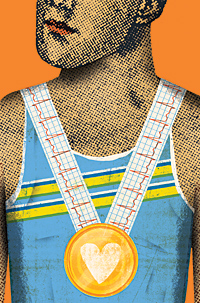
It’s a claim that challenges conventional wisdom in the United States: Screening young athletes for heart abnormalities is a cost-effective way to save lives. Stanford cardiologists came to the conclusion as a result of their study, published in the March 2 Annals of Internal Medicine.
Currently in the United States, routine screening to spot diseases that can cause sudden cardiac death is required for many professional athletes but not for those in college or high school. Participation requirements for these younger athletes are usually limited to physicals and medical history. In general, the thinking goes, heart testing — which uses electrocardiogram tests, or ECGs — is a good idea but it’s just too expensive, especially when the prevalence of death is so low.
But a study out of Italy, where routine ECG heart screenings of young athletes have been mandatory since 1982, has fueled the debate about the feasibility of such testing in the United States. The study showed that sudden death during competition has decreased nearly 90 percent since testing began.
The Stanford authors based their model on results from the Italian study, while adjusting for U.S. variations in disease prevalence and differences in screening modalities. They also collected data from numerous smaller studies written on the topic.
“We wanted to do a scientific analysis of cost-effectiveness,” says senior author Euan Ashley, MD, PhD, assistant professor of cardiovascular medicine and director of the Stanford Hypertrophic Cardiomyopathy Center.
Results showed that adding the ECG to screening at a total cost of $88 per athlete saves 2.1 life-years per 1,000 athletes screened. The total cost included initial testing, follow-up and treatment resulting from screening. From a health economics standpoint, the cost-effectiveness ratio was $42,900 per life-year saved.
“The cost-effectiveness ratio means you spend more money screening, but you also get benefits by reducing deaths,” explains co-author Mark Hlatky, MD, professor of health research and policy and of cardiovascular medicine. “Procedures under $50,000 per life-year added are generally accepted in the United States as cost-effective. Procedures over $100,000 are often not.”
Sudden death in athletes is rare. What researchers are looking for, when they examine the athletes’ ECG results, are signs of hypertrophic cardiomyopathy, long QT syndrome and other causes of sudden death in young athletes. In the United States, hypertrophic cardiomyopathy is the most common cause
of athlete death. An estimated one out of every 500 people in the nation has the condition, the first symptom of which can be death.
— Tracie White
The study was funded by the Breetwor Foundation and the Stanford Cardiovascular Institute.
MS: Divide and conquer
Christian Northeast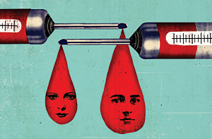
The discovery that there may be two distinct versions of multiple sclerosis has big implications for patients: Their responsiveness to the most popular first-line treatment seems to depend on which version they have.
If larger studies of patients confirm the findings, a simple blood test might someday tell people with multiple sclerosis whether they are likely to respond to the standard therapy, says senior study author Lawrence Steinman, MD, professor of neurology and neurological sciences.
Public health may benefit, too, Steinman says, as the cost savings from predicting which patients will benefit from beta-interferon, a costly bioengineered drug whose global sales come to some $4 billion a year, could be considerable.
In the study, published online March 28 in Nature Medicine, Steinman and his colleagues used an animal model of multiple sclerosis called experimental autoimmune encephalitis, or EAE, which prompts the immune system to inappropriately attack the animals’ own myelin nerve-cell coatings.
Many nerve cells in mammalian brains and peripheral tissues must convey electrochemical impulses over great distances, and quickly. Long, wirelike projections that transmit these cells’ signals to other nerve or muscle cells are coated by myelin, a natural substance whose insulating properties sustain the impulses’ strength and increase their speed.
Multiple sclerosis is triggered when, for reasons that are not clear, immune cells called T cells attack the myelin sheathing, causing symptoms including paralysis and blindness.
As for EAE, researchers can induce it using two specific chemicals that T cells secrete into the blood: gamma-interferon and IL-17.
In the new study, Steinman’s team induced two similar forms of EAE in mice by directing the myelin-attacking T cells to predominantly secrete either gamma-interferon or IL-17. The researchers found that beta-interferon improved the condition of animals whose EAE had been induced by gamma-interferon-secreting T cells, but exacerbated symptoms in those whose EAE had been induced by IL-17-secreting T cells.
Intrigued, the investigators turned to humans. The Stanford group obtained blood samples taken from 26 multiple-sclerosis patients who had been treated with beta-interferon. They measured IL-17 levels in those samples, and a clear pattern emerged. Measurements of a particular variety of IL-17, called IL-17F, clustered at either very high or very low levels in individual patients’ blood. Those with very low detectable blood levels of IL-17F responded well to beta-interferon treatment. But patients with very high IL-17F levels — about one out of three subjects — responded poorly. In fact, says Steinman, there is some evidence that beta-interferon actually worsened these patients’ conditions.
Steinman cautions that the results need to be confirmed in larger patient groups, but adds, “I think this has the potential to transform the way we take care of people with multiple sclerosis.” He says a simple, already available blood test could spare many patients the inconvenience and side effects of a drug that most likely won’t do any good. — Bruce Goldman
Stanford’s Office of Technology Licensing has filed a patent application on the use of the blood test described above. The research was funded by the National Multiple Sclerosis Society.
New neurons
Even Superman needed to retire to a phone booth for a quick change. But now scientists at the School of Medicine have succeeded in the ultimate switch: transforming skin cells into nerve cells, which are much harder to come by. And unexpectedly, the skin cells make the change without first becoming a stem cell — a step long thought to be required for cells to acquire new identities.
“We actively and directly induced one cell type to become a completely different cell type. These are fully functional neurons. They can do all the principal things that neurons in the brain do.”
The finding could revolutionize the future of human stem cell therapy and recast our understanding of how cells choose and maintain their specialties in the body.
“We actively and directly induced one cell type to become a completely different cell type,” says Marius Wernig, MD, assistant professor of pathology. “These are fully functional neurons. They can do all the principal things that neurons in the brain do.”
Wernig is the senior author of the research, published online Jan. 27 in Nature, which marked the first time that skin cells have been converted into fully functional neurons in a lab dish.
Until recently, it’s been thought that cellular specialization was a one-way path: Embryonic stem cells give rise to all the cell types in the body, but as the daughter cells become more specialized, they also become more biologically isolated. A skin cell could no more become a nerve cell than Superman could become Clark Kent in midair.
That view began to change when Dolly the sheep was cloned from an adult cell in 1997, showing that, under certain conditions, a specialized cell could shed these restrictions and act like an embryonic stem cell.
Old cells, new tricks
A decade later, researchers had made more progress in coaxing adult cells to act like embryonic ones: In 2007, they announced the creation of induced pluripotent stem cells, or iPS cells, from human skin cells by inserting four proteins called transcription factors into their DNA. The cells are called pluripotent because they have the ability, as do embryonic stem cells, to give rise to all other cell types.
Finally, in 2008, researchers showed it was possible in adult mice to reprogram one type of cell in the pancreas to become another pancreatic cell type by adding just three transcription factors.
As a result, Wernig began to wonder whether the pluripotent pit stop was truly necessary.
To test the theory, his team amassed a panel of 19 genes involved in either cell reprogramming or neural development and function. Then they inserted the genes into skin cells from embryonic mice and monitored the cells’ responses. After 32 days, some of the former skin cells looked like neural cells and expressed neural proteins.
The researchers winnowed the original pool of 19 genes down to just three, then tested the procedure on skin cells from the tails of adult mice. They found that about 20 percent of the former skin cells transformed into neural cells in less than a week — a vast improvement over the weeks-long iPS process in which only about 1 to 2 percent of the original cells become pluripotent.
“This is much more straightforward than going through iPS cells, and it’s likely to be a very viable alternative,” Wernig says.
Quickly making neurons from a specific patient may allow researchers to study particular disease processes, such as Parkinson’s, in a lab dish, or one day to even manufacture cells for therapy. — Krista Conger
The research was supported by Stanford’s Institute for Stem Cell Biology and Regenerative Medicine, the Donald E. and Delia B. Baxter Foundation, William Stinehart Jr. and the Reed Foundation, and the National Institutes of Health.