
SPRING 2010 CONTENTS
Home
Little patients, big medicine
Getting serious about helping without hurting
Girls’ day out
Teens with cancer paint the town pink
Hand-me-down blues
Ending depression’s legacy
Paging mom and dad
The future of children’s hospitals
Mother Courage
Mia Farrow’s calling
The inner child
Art offers an opening
A most mysterious organ
Looking for answers about the fetus’s lifeline

DOWNLOAD PRINTABLE
ISSUE (PDF)

Sugar time
Scientists have long struggled to understand the body’s biological clock. Its ticktock wakes us up, reminds us to eat and tells us when to go to bed. But what sets that circadian rhythm?
New research now shows that daily fluctuations in powerful hormones called glucocorticoids directly synchronize the biological clock by regulating “clock genes” that go on to play an important role in controlling blood sugar levels.
Christopher Silas Neal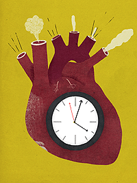
“The most surprising part of our findings is that our internal biologic rhythms are embedded directly into another pathway, one that is essential to regulate metabolism,” says the study’s senior author, Brian Feldman, MD, PhD, assistant professor of pediatric endocrinology at the medical school. Feldman also practices at Lucile Packard Children’s Hospital.
The findings give the first in vivo evidence of a direct link between glucocorticoid hormones and genes that regulate our biological clock. The study was published online Oct. 5, 2009, in Proceedings of the National Academy of Sciences.
Feldman’s team applied a synthetic glucocorticoid to dishes of mouse and human stem cells to see which genes responded. To the team’s surprise, three genes known to control the biological clock changed their activity in a direct response to the hormone.
Next, the researchers tested how the hormone’s effect on the biological clock is linked with its other functions. The scientists gave the synthetic glucocorticoid to genetically engineered mice lacking a specific gene involved in regulating biologic rhythms. As the team expected, genetically normal control mice responded with blood glucose changes associated with increased diabetes risk. In contrast, the genetically engineered mice were protected from harmful effects on blood sugar levels. The result shows that blood sugar regulation and the biological clock are entwined.
Timing’s everything
The close link between daily cycles of glucocorticoids, the body’s daily rhythms and blood sugar fluctuations should prompt doctors to examine how they use glucocorticoid drugs, Feldman says. For instance, prednisone is a powerful immune-suppressing glucocorticoid used to treat everything from severe asthma to cancer. Unfortunately, its side effects include poor regulation of blood sugar, weight gain and diabetes.
“Some very simple modifications in how we use glucocorticoids may change whether these drugs cause diabetes,” he says. Giving prednisone in a daily pattern that matches the body’s natural glucocorticoid cycle — with a peak in the early morning — might help solve the problem, he said. And because prednisone is already approved for human use, clinical trials of this idea would be fast and simple. — Erin Digitale
The research was funded by a grant from the National Institutes of Health.
Early cancer warning?
Christopher Silas Neal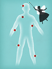
Imagine waving a magic wand over a region of a person’s body to determine if he has early signs of cancer. Researchers at the School of Medicine have shown in a laboratory study that ultrasound energy delivered to specific body regions can trigger the release of biochemical disease markers from tissues, making those markers more detectable as well as helping to pinpoint their source.
Upping the signal
Cancer cells produce substances not normally made by healthy cells and release those telltale biomarkers into the blood. In principle, this should allow relatively noninvasive early detection of tumors by monitoring patients’ blood for various cancer biomarkers. But only a handful of them are used in clinical practice.
Circulating levels of some biomarkers are ordinarily too small for reliable detection, but there’s a way around this. The cells that produce them carry multiple copies of these substances on their membranes — and at the right frequencies, ultrasound perturbs the membranes, spilling larger amounts of biomarkers into the bloodstream.
“Tumors are often not detected until they become sizable enough to cause recognizable symptoms or to be observed themselves as masses on an X-ray or CT scan,” says Gary Glazer, MD, professor and chair of radiology. “By then, the prognosis is much poorer than it might have been had the detection occurred earlier.”
Ultrasound energy delivered to specific body regions can trigger the release of biochemical disease markers from tissues, making those markers more detectable as well as helping to pinpoint their source.
In a study published online Sept. 21, 2009, in Proceedings of the National Academy of Sciences, Glazer’s team reported that they applied ultrasound to cultured colon cancer cells, causing the cells to release large amounts of a biomarker known as CEA into the surrounding culture medium. Then they optimized the ultrasonic energy level to trigger substantial CEA release from the cells without killing large numbers of them. (An important ultimate clinical objective is to be able to find a patient’s tumor without killing adjacent healthy tissue during the search.)
The researchers then implanted CEA-producing tumor cells into mice with impaired immune systems. Once the cells had engrafted to form tumors, ultrasound beams were directed at both tumor-bearing and, for comparison, tumor-free sites. Ultrasound energy aimed at tumors significantly increased the mice’s blood levels of CEA compared with the levels observed either before tumor cells had been implanted or when the beam was applied to non-cancerous sites. — Bruce Goldman
The study was funded by grants from the Lucas Brothers Foundation, National Institutes of Health, National Cancer Institute, Canary Foundation and Stanford’s Department of Radiology.
Kidney punch
Christopher Silas Neal
For older residents of U.S. nursing homes, starting dialysis may do more harm than good, heralding a decline in their ability to perform daily tasks such as feeding themselves, getting dressed or brushing their teeth, say researchers at the medical school.
Within the United States, 400,000 patients receive dialysis — a method of removing waste products from the blood when the kidneys fail. The treatment can be particularly burdensome for frail elderly people because they must travel to and from the dialysis centers, typically three times a week for three to four hours per treatment.
Americans over the age of 80 are the fastest-growing segment of the dialysis population, an increase not explained simply by population growth or an increase in diseases that cause kidney failure. This appears to be because physicians are much more willing to provide dialysis therapy to the very elderly, says Manjula Kurella Tamura, MD, assistant professor of nephrology. At least a third of these patients suffer from multiple chronic illnesses, such as heart disease and diabetes, in addition to kidney failure.
“The findings are sobering,” says Kurella Tamura, who conducted the study with colleagues at Stanford and UC-San Francisco. “One of the rationales for starting dialysis in patients with limited life expectancy due to diseases other than kidney failure is that, even if dialysis doesn’t extend life, it will improve the quality of life by alleviating symptoms of kidney failure or improving the ability of patients to care for themselves.”
Depending on a patient’s other medical problems, this may not be true, according to the study by Kurella Tamura and her colleagues in the Oct. 15, 2009, New England Journal of Medicine.
The study identified 3,702 nursing home patients from national registries who started dialysis between June 1998 and October 2000 and who had at least one measurement of their functional status available before they started the treatment. Functional status was measured by assessing the degree of dependence in seven activities of daily living.
Researchers then compared the patients’ functional status over the year prior to dialysis with the status over the year following treatment. Results showed that 12 months after starting dialysis, 58 percent of the patients had died, and only 13 percent had maintained the functional level they had before starting dialysis.
“We have tended to overestimate the benefits and ignore or downplay the negative aspects of dialysis when we counsel patients about their treatment options,” Kurella Tamura says. — Tracie White
The research was supported by the National Institute on Aging and grants from the National Center for Research Resources and the National Institute of Diabetes and Digestive and Kidney Diseases.
Rejecting rejection
The first doctors she consulted told Rachel Amato that the odds were vastly against her ever having a successful kidney transplant.
Then the 29-year-old mother of four from Turlock, Calif., traveled to Stanford Hospital & Clinics. “They were really honest with me,” she recalls. “They said, ‘We have seen proof that this new treatment works. We just don’t know how many doses it’s going to take.’”
In August 2008, Amato had her first four-hour infusion of a “desensitization” drug, intravenous immunoglobulin, or IVIG, designed to lower the number of organ-rejecting antibodies in patients who are highly “sensitized.” That means they have weapons-grade antibodies — acquired in blood transfusions, pregnancies or during a previous transplant — that seek out and destroy invading antigens, particularly the human leukocyte antigens, or HLA, on the surface of a foreign organ. These patients’ immune systems would reject a donor kidney.
Amato had one monthly infusion for four months, and in January 2009 she had a dose of an additional drug, Rituxan, which knocks out many of the immune-system cells that produce antibodies. “Boom! My antibodies dropped,” she says. “I got my transplant two weeks later.”
The desensitization program, as well as improvements in minimally invasive surgery and the promise of an experimental “tolerance induction” protocol, have placed Stanford at the forefront of kidney transplant programs. It was the only one among 240 kidney transplant centers nationwide that exceeded expected results in both patient and graft (transplant kidney) survival at one year and at three years after transplantation, according to the independent Scientific Registry of Transplant Recipients. The registry also shows that Stanford was the top program in one-year transplant kidney survival rates for four years running, from July 2000 to June 2004.
“It’s not just a fluke,” says Stephan Busque, MD, surgical director of the adult kidney and pancreas transplant program. “Even though we’re treating patients at higher risk, we perform better than expected because we have a very good team and our patients get very attentive, individualized care.”
Busque and the other members of the program have been pursuing new tests, treatments and technologies to address the plight of people who, like Amato, need transplants but can’t qualify for them because they are highly sensitized.
The new protocol involves giving these patients a high dose of IVIG. The infusions, which may be repeated over several months, lower the patient’s number of organ-rejecting antibodies.
Many could benefit
“They were really honest with me,” she recalls. “They said, ‘We have seen proof that this new treatment works. We just don’t know how many doses it’s going to take.’”
Dolly Tyan, PhD, director of the program’s histocompatability lab, estimates that 30 percent of the 80,000 people on the national waiting list are HLA sensitized and might benefit from being desensitized. The treatment is available at only a handful of medical centers, including Cedars-Sinai, Johns Hopkins, the Mayo Clinic and the University of Toronto.
Tyan and her colleagues have also developed a new assay system that “allows us to see very specifically what antibodies a person has, and exactly which ones are going to go away.”
In the meantime, Amato is grateful that she had the opportunity to get a kidney. While she still takes medication to ensure against her body rejecting the organ, her life is almost back to normal. “Stanford was my saving grace because they did not give up on me,” she says. — Diane Rogers
Sperm from scratch
“Figuring out the genetic ‘recipe’ needed to develop human germ cells in the laboratory will give us the tools we need to trace what’s going wrong for these people.”
Scientists have a promising new tool for understanding infertility: cells derived from unused embryos created for in vitro fertilization.
Stanford researchers have devised a way to efficiently coax stem cells derived from embryos to become germ cells, which are the precursors of egg and sperm cells. And unlike previous methods, which yielded primarily immature germ cells, the cells made with the new approach generated nearly mature sperm.
“This is the first evidence that you can create functional human germ cells in a laboratory,” says senior author Renee Reijo Pera, PhD, director of Stanford’s Center for Human Embryonic Stem Cell Research and Education.
“Ten to 15 percent of couples are infertile,” says Reijo Pera. “About half of these cases are due to an inability to make eggs or sperm. And yet deleting or increasing the expression of genes in the womb to understand why is unethical. Figuring out the genetic ‘recipe’ needed to develop human germ cells in the laboratory will give us the tools we need to trace what’s going wrong for these people.” The study was published online by Nature on Oct. 28, 2009.
Previous efforts to study infertility have been hampered by the fact that the human reproductive cycle cannot be adequately studied in animal models. And because germ cells begin to form very early in embryonic development (by eight to 10 weeks), there’s been a dearth of human material to work with.
Her team first isolated stem cells from the unused embryos, treated them with proteins that stimulate germ cell formation and isolated those that began to express germ-cell-specific genes — about 5 percent of the total.
They then used a technique called RNA silencing to examine how blocking the expression of each of three genes known to be involved in male infertility affected germ cell development. Conversely, they also investigated what happened when these genes were overexpressed.
They found that one of the genes functions very early in germ cell development, while two others stimulate the then-mature germ cells to form immature gametes, the next step toward becoming sperm and eggs. Overexpressing the three proteins together allowed the researchers to generate cells expressing proteins found in mature sperm.
After 14 days of this treatment, about 2 percent of the differentiated human embryonic stem cells had only one copy of each chromosome — indicating they were germ cells. (Most other human cells, including normal, untreated embryonic stem cells, have two copies of each.)
The researchers plan to use a similar strategy to optimize the production of eggs from embryonic stem cells, as well as investigating whether reprogrammed adult cells called induced pluripotent cells can also be used to create germ cells. By charting the germ cells’ development, they hope to identify problems that lead to infertility, miscarriage and birth defects. — Krista Conger
The research was funded by the National Institute of Health, the California Institute for Regenerative Medicine and the Tobacco-Related Disease Research Program.