Picture imperfect
See what Monet, Degas saw
By TRACIE WHITE
Michael Marmor, MD, wanted to know what it was like to see through the eyes of an artist.
Literally.
After writing two books on the topic of artists and eye disease, the Stanford ophthalmologist decided to go one step further and create images that would show how artists with eye disease actually saw their world and their canvases. Combining computer simulation with his own medical knowledge, Marmor has re-created images of some of the masterpieces of the French impressionistic painters Claude Monet and Edgar Degas who continued to work while they struggled with cataracts and retinal disease, respectively.
|
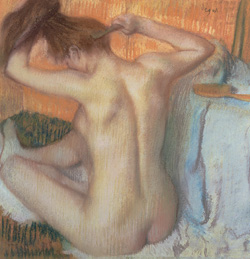
Hermitage, St. Petersburg/Bridgeman Art Library |
|
 |
 |
Edgar Degas completed this pastel titled Woman Combing Her Hair, in 1886. |
The results are striking.
In Marmor’s simulated versions of how the painters would most likely have seen their work, Degas’ later paintings of nude bathers become so blurry it’s difficult to see any of the artist’s brushstrokes. Monet’s later paintings of the lily pond and the Japanese bridge at Giverny, when adjusted to reflect the typical symptoms of cataracts, appear dark and muddied. The artist’s signature vibrant colors are muted, replaced by browns and yellows.
“These simulations may lead one to question whether the artists intended these late works to look exactly as they do,” says Marmor who has long had interest in both the mechanics of vision and vision in artists. “The fact is that these artists weren’t painting in this manner totally for artistic reasons.”
Degas and Monet were both founders of the Impressionist era, and the style of both painters was well-formed before their eye disease affected their vision. But their paintings grew significantly more abstract in later life as their eye problems increased.
“Contemporaries of both have noted that their late works were strangely coarse or garish and seemed out of character to the finer works that these artists had produced over the years,” Marmor writes in a paper titled “Ophthalmology and Art: Simulation of Monet’s cataracts and Degas’ retinal disease,” which was published in the December issue of the Archives of Ophthalmology.
It’s well-known that such artists as Monet, Degas, Rembrandt, Mary Cassatt and Georgia O’Keefe all reached their heights of artistic vision while facing a decline in their ocular vision. Marmor chose to focus on Degas and Monet because both artists suffered from eye disease that was documented in historical records, journals and medical histories. Degas had retinal eye disease that frustrated him for the last 50 years of his long career. Monet complained of cataracts interfering with his ability to see colors for 10 years before he finally underwent surgery to have them removed.
|
Norton Simon Art Foundation |
|
 |
|
By the time Degas completed Woman Drying Her Hair in 1905, his eyesight had dropped to somewhere between 20/200 and 20/400. Marmor notes that after 1900, there was virtually no detail in faces or clothing in Degas' artwork. |
“We understand better from these simulations what Degas and Monet struggled with as vision failed,” Marmor says.
Over the past 32 years, the Harvard-educated physician has published 200-plus scientific articles on the science of eye disease while at the same time writing about famous artists and how eye disease may have affected their artwork. He authored one book, Degas Through His Own Eyes, and co-authored another, The Eye of the Artist, with ophthalmologist James G. Ravin, MD.
“As an ophthalmologist, I’m fascinated with the visual components of art,” says Marmor, whose Stanford home is decorated with pieces of modern art that emphasize optical illusions. His family donated works of art to the Cantor Arts Center at Stanford. “I’ve also spent years talking to patients about the symptoms of their eye diseases. This was a natural outgrowth of my science and art interests.”
Jody Maxmin, PhD, Stanford associate professor of art and art history and of classics, says that Marmor’s work is compelling to historians of ancient art.
“Dr. Marmor’s research inspires us to imagine how aging artists must have grappled as their vision changed,” Maxmin says. “It inspires us to look at the topography and monuments of the ancient world with ‘ancient’ eyes, eyes that would have been sharp and reliable for only so long.”
|
Michael Marmor |
|
 |
|
Marmor blurred the image in Woman Drying Her Hair to a visual acuity of 20/300 to replicate what Degas might have seen.
|
To create the images of the artists’ paintings as seen through their own eyes, Marmor used Adobe Photoshop software for photo editing. He adjusted the blur and filter settings to what he determined would be the different stages of Degas’ and Monet’s eye disease, based on medical expertise and historical research.
Degas suffered failing vision from 1860 to 1910. As his eye disease progressed, his paintings grew increasingly rough. From treating hundreds of patients with retinal disease similar to what Degas suffered, Marmor says, he knows that the shading and contrast of images becomes less defined and blurriness increases as such illness progresses.
“Friends would ask Degas, ‘Why are you still painting?’” Marmor wrote in his December paper. “His works in the 1870s were drawn quite precisely with facial detail, careful shading and attention to the folding of ballet costumes and towels.’’ By the 1880s and 1890s, the shading lines and details of the face, hair and clothing of the same subjects became progressively less refined.
“After 1900,’’ Marmor says, “these effects were quite extreme and many pictures seem mere shadows of his customary style.”
Monet wrote of his growing frustration with his deteriorating vision, describing how he was forced to memorize where the colors were placed on his palate. In 1914 he wrote in his correspondence that colors no longer had the same intensity. “Reds had begun to look muddy,’’ he wrote. “My painting was getting more and more darkened.” He was forced to rely on the labels on the tubes of paint in place of his own vision.
“Like retinal disease, cataracts also blur vision,” Marmor says, “but more importantly for a painter like Monet, whose style was based on the use of light and color, they can affect the ability to see colors.”
Marmor wrote in the 2006 paper that, “Monet must have struggled mightily as he looked out into the murky yellow-brown garden and tried to decide what subtle impression to create on canvas. Slowly progressive age-related cataracts manifest as yellowing and darkening of the lens. This has a major effect on color perception as well as visual acuity.”
After reluctantly submitting to cataract surgery in 1923, Monet returned to his original painting style, even throwing away much of the artwork he’d done during the 10-year period that he had cataracts.
“He just couldn’t see the colors,” Marmor says. “These simulations show how much his sense of color had been destroyed. Some people say, ‘Oh, it’s a stylistic change.’ Gosh, I don’t think so.”
Understanding the challenges these artists faced because of eye disease helps further illuminate the accomplishments they achieved despite their disabilities, Marmor says.
“There’s some reluctance among people in the art world to look outside the historical or psychological influences on the great artists,” Marmor says. “I’m open to debate about what these visual changes might mean stylistically or aesthetically. What is not open to debate is what the artists saw. If you ignore that, you’re ignoring facts.”
Comments? Contact Stanford Medicine at
 Back
To Contents
Back
To Contents