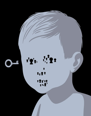
About face
The secret of your smile remains one of life's persistent questions
By KRISTA CONGER
In 1995, a class of Minnesota school children on a field trip to a local farm pond collected some unusual inhabitants: frogs with extra or missing limbs. Their discovery, followed in 1996 by a rash of deformed frogs along the shores of Vermont’s Lake Champlain, ignited a wave of concern about possible contaminants in the mud and water in which the frogs lived. A U.S. Geological Survey study found hundreds of similarly malformed frogs across the northern states and California. A few ended up in the hands of Jill Helms, DDS, PhD, who promptly trained her microscope on the frogs’… heads.
Helms, a specialist in craniofacial development and an associate professor at Stanford’s School of Medicine, had a hunch that the limb-challenged frogs’ problems didn’t stop at the neck. She was right. The frogs also had other, more subtle defects in their skulls and faces mimicking the lip and palate clefting that, at a frequency of about one in every 500 to 600 births, is the most common birth defect in humans.
It’s more than an environmental issue. Understanding the how and the why of facial formation is the first step to preventing, diagnosing and perhaps even treating not just human birth defects, but also damage caused by trauma or disease. Ideally Helms, who is supported by the Lucile Packard Foundation for Children’s Health, and her colleagues will one day be able to knit bones and mend skin with the flip of a molecular switch — transforming lives in a world where appearances matter almost as much as function.
Facial anomalies are so common in part because the process of forming faces is so complex. Environmental contaminants, maternal alcohol or medication use, or even a random glitch in protein production can all short-circuit the molecular dance necessary to craft a nose, eyes and ears from a mass of undifferentiated cells. Studying the hapless frogs is just one way to shed some light on the little understood process.
One way to investigate how faces form is through the time-honored method of tinkering. In 2003 Helms, then at UC-San Francisco, showed that she could create “quck” and “duail” embryos — that is, quails with duck beaks and ducks with quail beaks — by simply transplanting pinhead-sized bits of specialized tissue from one species to another early in development. Although this experiment showed that some cells clearly have ingrained marching orders that both direct and limit what they can become, other experiments indicated that neighboring cells play a large part in determining a cell’s fate. The conflicting results had many scientists scratching their heads.
“There’s a fair amount of hand waving when it comes to describing facial development,” says Helms. “But what we do know is that there’s zero margin for error.” Any disruption can cause a range of malformations from a barely noticeable cleft lip to a fatal form of a condition called holoprosencephaly that leaves infants with only one eye in the center of their forehead, one central tooth and little or no separation between the two hemispheres of the brain. Other developmental missteps can result in noses that are grossly misshapen or absent altogether.
Faces on the brain
Helms didn’t start her scientific career interested in faces. An ambitious neurobiology graduate student with a dental degree, she wanted to understand how the brain develops. To gain the technical skills necessary to understand the basic mechanisms by which patterning and growth are regulated, she signed on as a post-doc in Gregor Eichele’s laboratory at the Baylor College of Medicine, which at that time focused on limb development. But her interest was piqued when she noticed that many of the signaling molecules orchestrating the growth of legs and wings were also expressed in the animal’s head and face — a fact that was ignored by her limb-centric colleagues.
“When I asked my colleague Cliff Tabin if he’d ever noticed, he said, ‘Jill, the heads are just the handles by which we hold the embryos,’ ” says Helms. “Unlike limbs, which are so nice to study — they have only a couple of bones, they stick out conveniently from the body — the face, oh my god! There’s active migration, paths of displacement and neural development all going on simultaneously. There’s a lot of movement going on at that end of the embryo.”
But Helms was undeterred, and by the time she was at UCSF her qucks and duails were gaining worldwide attention. It was clear she’d gone over to the wrong side of the embryo when she started referring to everything below the neck as the “peri-anal region” — a not-so-subtle tease of her former colleagues.
 |
|
|
|
|
|
|
|
|
|
|
|
|
|
|
|
|
|
What’s in a face
Untold numbers of songs and poems have been written about the human heart. But it’s the face that’s the true workhorse in our interactions with the outside world. Not only does it house four of our five senses, it’s also the main way we express our feelings — intentionally or unintentionally — to those around us. It’s so important that there is at least one chunk of the brain, the fusiform gyrus, devoted simply to recognizing faces, and another to deciphering their meaning.
Paul Ekman, PhD, found through studies of isolated tribes in New Guinea in the 1960s that facial expressions are universal — happy people look happy regardless of what part of the world they live in and what type of culture they grew up in. Ekman spent the next several decades meticulously identifying groups of “microexpressions” that, when correctly interpreted, together belie a person’s true feelings regardless of what he or she might be saying or doing.
“I have not been able to find an emotional expression that isn’t universal,” says Ekman, a professor of psychology at UC-San Francisco, in a 2004 video interview for UC-Berkeley’s Conversations with History series. His technique is so effective it’s taught to Secret Service agents and the FBI to give an edge to professionals paid to suss out the truth.
Since a face conveys so much meaning, deformities result in great psychological pain. In addition to any functional problems that may exist, the individual is uniquely cut off from society by an inability to communicate with easily understood facial expressions and an almost instinctive reluctance of others to try to interpret unfamiliar facial cues.
The face is such an effective communication tool that it’s often seen as the repository of someone’s personality, or “self.” Such confusion might be why the face transplants recently reported in the worldwide media hold such fascination. “There’s a perception that the dead person’s personality will also be transferred to the recipient,” says craniofacial development researcher Jill Helms, DDS, PhD, “kind of like how it seemed that the first heart transplants might also be bestowing some part of the donor’s soul.”
The importance of faces, the devastation of facial deformity and the reason researchers like Helms, a professor of surgery at Stanford, are trying so hard to correct abnormalities, might be best reflected by an experiment in which very young children were asked to identify pictures of body parts. “That’s a leg,” they would say appropriately. “That’s an arm, that’s a foot,” they would continue, but — when shown a picture of their face — “Oh! That’s me.” |
|
|
|
|
|
|
|
|
|
|
|
|
|
|
|
|
|
|
Why should a human who possesses neither a duck’s bill nor a bird’s beak care about whether this structure is pointy or rounded? Most vertebrates look pretty much the same during early development and use similar pathways to create very distinctive looks.
“If we’re trying to figure out what causes facial variation in humans, it makes sense to study it in birds,” says Helms. We would never mistake a mouse for a bird, or a bird for a human, but in the very early stages of embryonic development the head regions look identical. Within a few short days, however, the differences between the species are fully evident: a result of variations in local proliferation that drive some primordial tissues out and cause others to shrink, modulating appearances.
Recipe for a nose
But there’s quite a bit happening backstage in vertebrates even before this bit of fine tuning. The cells that make up structures of the face, including the nose, originate from the nascent central nervous system. Early in mouse development, after gastrulation forms the digestive tube, the neural tube forms along what is to be the back of the embryo, first as a thickened layer of cells and then as a shallow trough between two growing waves of tissue. After six to seven days, the waves meet at one or more locations along the top and begin to “zip” closed in both directions, leaving a border of cells known as the neural crest.
The neural crest cells at the head end of the embryo have a daunting task. Like Dorothy in The Wizard of Oz, they must make their way through an unfamiliar landscape, following a yellow brick road of signals generated by wayside cells in order to wend their way from the back of the embryo around to the facial primordia. Once there, they strike a delicate balance between following sage advice from nearby developing brain cells and staying true to their internal cues as they differentiate into their final form.
Most of the time they get it right. Sometimes they don’t. Many researchers conducting transplantation experiments believe that once the number of non-native neural crest cells reach a critical mass — think the Tin Man, the Cowardly Lion and the Scarecrow — they’re less likely to assimilate into their new location. Hence, qucks and duails. In addition, Helms has found that interfering with key signaling pathways, such as those mediated by the proteins Wnt and Sonic hedgehog, can muck up both brain and face development in a time-dependent way. For holoprosencephaly, earlier interference with the Sonic hedgehog pathway causes more severe defects than blocking the signal later in development.
“We’re finding that individual pathways function throughout the body like a web,” says Helms, “and that limb and facial development use many of the same genes. In fact, there is a huge group of birth defects characterized by abnormal limbs and faces. Now we understand how that could occur.”
Mice and dogs and pigeons, oh my
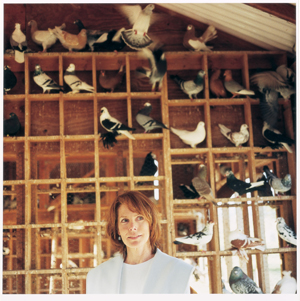
One of the difficulties of studying naturally occurring facial variation is finding an appropriate animal model. Even the most ardent rodent lover would have to concede that laboratory mice all pretty much look the same. In contrast, humans’ astonishing repertoire of noses, eyes, eyebrows and mouths makes it possible to categorize different ethnic groups.
“We really need a system in which one species exhibits enormous variation in facial structure,” says Helms. “Dogs spring to mind immediately. There’s the bulldog, the borzoi, the Saint Bernard. But dogs can be difficult laboratory animals to study.” For example, dogs require more space and have much longer gestation periods than mice. Although she’d had some success comparing ducks, chickens and quails, the differences between the species were potential stumbling blocks.
Finally she decided to take a cue from Darwin. But instead of going to the Galapagos for finches, she’s going to Gilroy for pigeons. It’s not as crazy as it might seem at first. Darwin himself spent years after returning to England studying and breeding pigeons to hone his evolutionary theory. The DeCarlo family of Gilroy, which has bred pigeons for three generations and maintains a large flock representing more than 40 breeds, gives Helms the same opportunity. Their meticulous breeding records document decades of producing birds that are vastly different from the common city dweller we all know.
“Humans have been breeding pigeons for thousands of years,” says Helms. “There are references to tame pigeons in Egyptian tombs.” And, she points out, every breeder seems to have his or her favorite pigeon.
“The sheer variety of beaks alone is astounding,” says Helms, sounding like a kid in a candy store as she describes pigeons with big, curved beaks eight or nine times larger than normal, pigeons with short owl-like beaks and pigeons with beaks so tiny they’re barely visible. “There are beaks with enormous wattles, long pointy beaks and little tiny hooked beaks,” she says. “I mean, who would have thought! They are going to be the ideal animal model to study variations that come about by natural selection.”
Looking forward
Under all this talk of birds and beaks are real medical problems with few answers. Currently the only treatment for birth defects like holoprosencephaly and cleft lips and palates is palliative at best. Surgery can sometimes help a mildly affected child look and function more normally, but severe abnormalities can be life threatening. The long-term goal of research like Helms’ is to find ways to diagnose craniofacial abnormalities early enough during gestation to allow effective intervention. Similar techniques might one day be used to correct damage to the face and skull caused by trauma or disease.
“One of the most touching moments of my career came when I was touring a Shriner’s burn ward,” says Helms. “I saw a mother and father waiting by the side of their 8-year-old son, who had been severely burned on his birthday. They looked at me as if to say ‘You have answers. You can help our child.’ That’s a very motivating thing. You want to work hard and work fast because there’s someone out there waiting for that answer.”
Asked if she thinks her work is off the beaten path, she pauses, and laughs. “Anybody who thinks that a two-snouted pig with three eyes is great,” says Helms, “well, I suppose that kind of puts you on the fringe.”
Comments? Contact Stanford Medicine at
 Back
To Contents
Back
To Contents