 |
|||
| Stanford Medicine |
| Current Issue |
| Contact Us |
| ALL Issues |
| 2008 Spring |
| 2007 Fall |
| 2007 Summer |
| 2007 Spring |
| 2006 Fall |
| 2006 Summer |
| 2006 Spring |
| 2005 Fall |
| 2005 Summer |
| 2005 Winter |
| 2004 Fall |
| 2004 Spring |
| 2003 Fall |
| 2003 Summer |
| 2003 Winter |
| 2002 Fall |
| 2002 Summer |
| 2002 Winter |
| 2001 Fall |
| 2001 Winter/Spring |
| 2000 Fall |
| 2000 Spring |
| 1999/2000 Winter |
| 1999 Summer |
| 1999 Spring |
A QUICK LOOK at the latest developments from Stanford University Medical Center
![]() Start your engines
Start your engines
![]() Tackling the stickleback
Tackling the stickleback
![]() Heart to heart
Heart to heart
![]() Mom's magic formula
Mom's magic formula
![]() Take a bow
Take a bow
![]() Real-world medicine
Real-world medicine
![]() Let there be (ultraviolet) light
Let there be (ultraviolet) light
Start your engines
Taking a good look under the hood of a special type of molecular motorcar is giving biochemistry professor James Spudich, PhD, insights into new designs for drugs. Spudich says every cell possesses a "dynamic city plan" that consists of molecular highways, construction crews, street signs, cars, fuel and exhaust. Molecular motors help control the cell's activities -- and could represent ideal drug targets.
Illustration: Noah
Woods |
|
 |
|
This spring, Spudich, who has long studied molecular motors, and physics graduate student David M. Altman revealed how one particularly important motor works.
In the March 5, 2004, issue of Cell, the pair show that a tension-sensitive molecular motor provides the rigidity needed by the tiny, hairlike sensors of the inner ear to respond to sound.
This study demonstrated for the first time how a protein could function as an anchor, stalling in place once the optimal tautness is created in a membrane and then restarting when the membrane slackens.
"Our general feeling is that tension-sensitive machines are at the heart of the dynamic city plan," says Spudich, who is also the Douglass M. and Nola Leishman Professor of Cardiovascular Disease.
With an understanding of the intricacies of the motors, researchers can design drugs to block or augment them; Spudich has co-founded a company called Cytokinetics to create drugs selectively targeting molecular motors involved in cancer and cardiovascular disease.
Hit the brakes
For example, some chemotherapeutic agents target rapidly dividing cancer cells by blocking the molecular highways -- called microtubules -- that are used by kinesins, motors critical to cell division. Microtubules are also used by nerve cells to relay information, so drugs that block these highways can cause profound neurological side effects.
It would make a lot more sense, says Spudich, to halt the specific motors involved in cell division rather than obstruct the highways along which they move. "If you have yellow Volkswagens running between San Francisco and Palo Alto on Highway 280 causing a problem you would like to inhibit, would you find some way to specifically blow up only the yellow Volkswagens traveling on that highway or would you blow up all the highways in the United States?" he says. "The answer is simple."-- Mitzi Baker
The study was funded by the National Institutes of Health.
|
![]() Back to Top |
Back to Top |
Tackling the stickleback
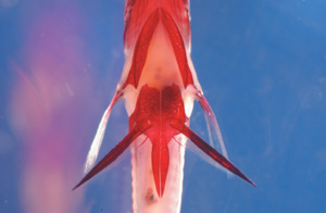
A small fish called the threespine stickleback is making big waves with researchers investigating how vertebrates evolve new traits. In an effort to move those studies forward, the National Institutes of Health recently added the stickleback to a list that includes cows, dogs, honeybees and other species that are all on the fast-track to having their genome sequenced.
Photo:
Michael Shapiro |
|
 |
|
| This threespine stickleback fish (from below) bears spiny pelvic fins. | |
David Kingsley, PhD, professor of developmental biology, first proposed sequencing the stickleback to the NIH in 2003. "The genome sequence of sticklebacks will make it possible to go from traits to genes much more quickly," he says.
Pockets of sticklebacks were isolated by geologic changes at the end of the Ice Age 10,000 years ago, with each newly separated population evolving in response to local ecological conditions. Although these populations now look distinct, in some cases only a few gene changes account for these differences.
Informative fish
Kingsley is among those who have made use of the threespine stickleback's unusual biology. He and his co-workers have caught hundreds of the minnow-sized fish and have bred the different populations to study their genetics. In a study published in the April 15, 2004, issue of Nature, he and his co-workers reported that two populations of sticklebacks lacking hind fins -- one from British Columbia and the other from Iceland -- carried a mutation in the same gene.
"People have been interested in limbs for a long time because they show such variability in different animals," Kingsley says. "The debate has been how many genes account for these differences."
The single gene mutated in these two fish populations is the stickleback version of a gene in mice called Pitx1 that, when mutated, causes mice to have greatly reduced hind limbs. "It looks like evolution is using this gene repeatedly," Kingsley says.
The NIH decision to fast-track sequencing of the fish's genome is great news, says Kingsley. Studies like his will go much faster with the sequence available. Sequencing will begin this year.
Kingsley serves on the NIH board that decides which species should be sequenced. Stanford geneticists Richard Myers, PhD, and Arend Sidow, PhD, also serve. Since all three have an interest in sequencing the fish, they bowed out of the stickleback discussion so no conflict of interest would influence the decision.
"With the sequence, we can begin using the system to determine the genetic and molecular basis of a broad range of traits that have evolved in different populations around the world," says Kingsley. -- Amy Adams
David Kingsley is a Howard Hughes Medical Institute Associate
Investigator. This work was supported in part by grants from HHMI; the
National Institutes of Health; the Natural Sciences and Engineering Research
Council of Canada; the Canada Foundation for Innovation; the City of Gardabaer,
Iceland; and the Helen Hay Whitney Foundation.
|
![]() Back to Top |
Back to Top |
Heart to heart
Imagine lying in a hospital bed, knowing that you have 24 hours to
live and your
sole hope for survival is a device that has been tested only in sheep.
Twenty years ago, that was the predicament facing Robert St. Laurent at
Stanford Hospital. He became the first successful human recipient of a
left ventricular assist device, or LVAD, which bought him eight days of
life -- long enough to receive a heart transplant.
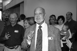
Photo:
Stanford Visual Art Services |
|
 |
|
| Robert St. Laurent at Stanford's annual transplant reunion. His historic LVAD surgery took place 20 years ago. | |
Last April, the 71-year-old St. Laurent traveled from his Florida home to Stanford's annual transplant reunion to join nearly 100 others who had received new hearts or lungs at Stanford. About 50 patients undergo heart transplantation each year at Stanford -- the most of any hospital in Northern California.
St. Laurent, one of the longest-living heart transplant survivors, "just happened to be in the right place at the right time," says Peer Portner, MD, consulting professor of cardiothoracic surgery, who developed the device used in St. Laurent. Portner's LVAD received Food and Drug Administration approval two months before St. Laurent arrived at Stanford in 1984.
Because the LVAD was untested in humans, the only information St. Laurent had to allay his apprehension was a report that the three sheep implanted with the device were going about their business, calmly lying down and chewing their cud, seemingly unconcerned.
Cynthia St. Laurent, Robert's wife, laughed so hard at this unconventional "patient update" that she spewed coffee all over her pink angora sweater.
A little more than a week after the LVAD was implanted by Philip Oyer, MD, the Roy B. Cohn-Theodore A. Falasco Professor of Cardiothoracic Surgery, St. Laurent received the heart of an 18-year-old college student who was left brain dead after a car accident -- a poignant twist for St. Laurent, who had a son the same age.
Transplant experts
Stanford has pioneered several advances in heart and lung transplantations, beginning with Norman Shumway, MD, the Frances and Charles Field Professor of Cardiovascular Surgery emeritus, and his colleagues performing in 1968 the first adult heart transplant in the United States. Bruce Reitz, MD, the Norman E. Shumway Professor of Cardiovascular Surgery, and his team successfully transplanted a heart and lung for the first time in the world in 1981.
With more than 1,000 heart transplantations under their belts, and more than 100 each of lung and heart-lung transplantations, Stanford's cardiothoracic surgery team is creating an ever-growing group of survivors with a new lease on life. -- Mitzi Baker
|
![]() Back to Top |
Back to Top |
Mom's magic formula
"Got breast milk?" This unconventional riff on the familiar dairy-industry ad is gaining popularity in the neonatal intensive care unit at Lucile Packard Children's Hospital. Why? Premature babies who receive breast milk leave the hospital about two weeks earlier than those who are formula-fed and are less likely to endure life-threatening infections or bowel disease.
Illustration: Noah
Woods |
|
 |
|
"Providing breast milk is one vital connection between mother and child that can be more useful than many of the medical procedures we can offer the baby," says Jane Morton, MD, director of the Breastfeeding Medicine Program at Packard Children's Hospital.
Because many premature babies are too small, weak or uncoordinated to nurse effectively, however, moms must use breast pumps to extract the milk for their infants. While this works well for the short term, it's difficult to trick the body into producing as much milk for a mechanical pump as it would have for a suckling baby.
"Some mothers are pump-dependent for months," Morton says. "However, because milk production plateaus after the first two weeks, it's really important to ramp up quickly even though the baby is still very small."
Establishing a high volume of milk early is critical to allow the baby to eventually switch from feeding tube or bottle to the breast, Morton says. "Without that volume the milk is too difficult for the baby to extract, setting up the mothers for early termination of breastfeeding." Tired moms who must offer their babies a bottle after first trying to breastfeed may not have the stamina to follow up with a pumping session to empty their breasts and stimulate more milk production.
Morton recently received a grant from Medela Corp. to compare the volume of milk produced by mothers using one of two pump models: one with a special "pre-letdown" setting and one with only the standard extraction pattern. The hope is that the pump tailored to stimulate letdown -- a physiological response to suckling that allows complete emptying of the breast -- will enhance overall milk production.
She'll also collect breast milk samples from women in the study to track changes in 20 common components. "We're interested in how the milk may vary between moms whose babies were born at different gestational ages. We know that breast milk from womenwho deliver prematurely tends to have higher protein and salt content, and it will be interesting to see how these and other levels may change as the baby ages." -- Krista Conger
|
![]() Back to Top |
Back to Top |
Take a bow
Springtime brought a shower of national and international accolades for medical school faculty members. In particular, three faculty were honored with some of the scientific world's most high-profile prizes.
Stanley Cohen, MD, the Kwoh-Ting Li Professor in the School of Medicine, received the 2004 Albany Medical Center Prize in Medicine, one of medicine's most prestigious awards.
He shared the $500,000 prize with Herbert Boyer, PhD, a founder of Genentech and professor emeritus at UC-San Francisco, for research that laid the groundwork for modern genetic engineering.
The pair's discovery allowed researchers to transfer pieces of DNA between organisms, a process also called DNA cloning, opening the door to the modern field of genetic engineering. Among the many lifesaving discoveries developed using DNA cloning are insulin to regulate blood sugar in diabetics, a clot-dissolving agent for stroke and heart attack victims, a human growth hormone for underdeveloped children and interferon for cancer patients.
The pair are also recipients of the newest million-dollar science award, the Shaw Prize. They share the Prize in Life Sciences and Medicine with UCSF professor Kan Yuet-Wai, MD. Hong Kong media executive Run Run Shaw, who made his fortune producing kung-fu action movies, established the prize in 2002.
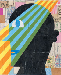
William Newsome, PhD, professor of neurobiology, won the 2004 Dan David Prize for his research into how the brain interprets visual signals. He shared the "Future" portion of the $1 million prize with two other neuroscientists. Additional awards were given in "Past" and "Present" categories.
Newsome was recognized for his research in primates on specialized brain cells that detect objects as they move across the field of vision. These are the cells that notice a fly buzzing from left to right or a bird taking off in flight. One of his most remarkable discoveries was reported in a 1990 Nature paper in which he and his colleagues showed that by artificially stimulating these neurons, a monkey would detect movement that wasn't there.
Eugene Butcher, MD, professor of pathology, received the $500,000 Crafoord Prize from the Royal Swedish Academy of Sciences, the same group that delivers the Nobel Prize. He split the award with Harvard biologist Timothy Springer, PhD.
Butcher was honored for his immunology work and its application to understanding arthritis. A decade ago, Butcher speculated that a regimented series of events was responsible for controlling how white blood cells, the immune system defense team that responds to an injury or infection, report for duty where they are needed. His theory is now widely accepted throughout the medical world and is being applied to the development of possible treatments for autoimmune diseases, such as arthritis. -- Susan Ipaktchian
|
![]() Back to Top |
Back to Top |
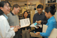
Real-world medicine
It's one thing to read about the plight of Americans who have little or no health insurance. But as third-year medical student Lyen Huang has discovered, it's quite another to look into the eyes of men and women who can't afford routine medical treatment.
Photo:
Steve Fisch |
|
 |
|
| A staff discussion at the Stanford medical student-run pacific free clinic in San Jose. | |
Huang is one of the managers of the medical student-run Pacific Free Clinic, which opened in May 2003. The clinic operates out of Overfelt High School in San Jose on Saturdays, providing free basic health-care services for low-income adults, most of whom are Chinese, Hispanic or Vietnamese.
About 94 percent of the clinic's patients lack insurance and are between the ages of 18 and 65, making them ineligible for Medicare or other types of health-care assistance.
"We know the clinic is a stop-gap measure," Huang says. "We can't see all of the patients who need our services, but we see it as a way to help connect the patients with other resources in the community."
Volunteer physicians, undergraduates and community members join the students in staffing the clinic.
Operating the clinic taught the students about the gaps in the U.S. health-care system. The students initially assumed they would mostly be treating one-time complaints -- colds, flu, rashes -- and conducting routine checkups and screenings. But it became clear that most patients needed treatment for chronic problems, such as diabetes and high blood pressure.
"They may have lost their job and can no longer afford their blood-pressure medication, so they come to the clinic complaining of headaches caused by high blood pressure," Huang says. "We could just treat the headache, but that doesn't solve the underlying problem."
The students opted to treat patients with chronic conditions, even though it meant potentially limiting the number of people the clinic could serve. In addition to providing medications, the clinic refers patients to other health-care programs in Santa Clara County that base their fees on a patient's income and are better suited to providing longterm care for those with chronic conditions.
With the clinic now in its second year of operation, the students plan to continue reaching out to community groups and searching for additional resources. "We would like the community to guide us," Huang says. -- Susan Ipaktchian
|
![]() Back to Top |
Back to Top |
Let there be (ultraviolet) light
It's possible a trip to the tanning salon might help patients with blood disorders, such as leukemia or lymphoma, who are about to undergo a bone marrow or stem cell transplant.
Illustration: Noah
Woods |
|
 |
|
At least that's the suggestion of a recent Stanford study in animals on graft-versus-host disease, or GVHD -- the condition that often causes these transplants to fail.
In GVHD, the newly transplanted cells attack the patient they're meant to help. The disease often appears as a reddish rash or in more severe cases, as skin that blisters and peels.
Stanford researchers believe they've found the disease's cause, an immune cell known as a Langerhans cell. These cells normally signal to T cells to fight off infection. But in transplants, they do patients a disservice, alerting T cells to attack the patient's own tissues, the researchers found.
How to aid transplants
The researchers were able to effectively eliminate the Langerhans cells in transplanted mice by using a sunlamp to expose them to ultraviolet light. The animals got a mild sunburn but suffered no other ill effects. Transplanted mice who got the sun treatment experienced no skin GVHD, while those that didn't showed severe signs of the disease, says Edgar Engleman, MD, professor of pathology and of medicine and senior author of the study. It was published in the May 2004 issue of Nature Medicine.
"The experiment not only provided the proof that these cells cause graft-versus-host disease but also provides a way of thinking about how you might prevent it," says Engleman, who directs the Stanford Blood Center.
Miriam Merad, MD, PhD, a former postdoctoral scholar in Engleman's lab and the study's first author, says the results could serve as the basis for a clinical trial in which patients were xposed to ultraviolet light before treatment. The trick would be to administer the ultraviolet treatment and deplete the Langerhans cells in a way that would avoid other, unwanted side effects, Engleman says. -- Ruthann Richter
This study was funded by the National Heart, Lung and Blood Institute and by a grant from the Irene Diamond Fund.
|
![]() Back to Top |
Back to Top |
Comments? Contact Stanford Medicine at